Sub-wavelength focusing of mid-IR light using metal/diamond/metal campanile probe for ultra-broadband SPM
- Abstract number
- 361
- Presentation Form
- Poster
- DOI
- 10.22443/rms.mmc2023.361
- Corresponding Email
- [email protected]
- Session
- Poster Session Two
- Authors
- Dr. Rajasekhar Medapalli (1), Dr. Khushboo Agarwal (1), Mr. Sergio Gonzalez Munoz (1), Dr. Sergey Kafanov (1), Dr. Samuel Jarvis (1), Dr. Rostislav Mikhaylovskiy (1), Prof. Oleg Kolosov (1)
- Affiliations
-
1. Department of Physics, Lancaster University
- Keywords
Mid-IR spectroscopy, Terahertz Spectroscopy, Advanced SPM
- Abstract text
Developing methods for efficient nanoscale probing of light-matter interaction, especially in the Mid-IR and THz spectral range, is essential for studying fundamental physical phenomena as well as chemical properties at micrometer to nanometer length scales. A highly efficient nanoscale focusing of visible and near-IR radiation light was reported recently using Au-SiO2-Au tapered gap campanile plasmon waveguide with an 830 nm wavelength that couples free space light into the nanoscale domain, enabling probing of materials in the visible and near-IR spectral range [1]. We expand this capability to the highly important mid-IR and THz range providing valuable information on local nanoscale chemistry and physical processes of materials and devices using a campanile shaped diamond tetragonal pyramid [2]. Our finite difference time domain (FDTD) simulation reveals that nanoscale focusing of mid-IR light is possible within the range of geometries and metal coatings including Au, Al and Cu. Here we report linked modeling and experimental results showing the confining efficiency of diamond pyramid in the mid-IR range (8-10 µm). Furthermore, we will demonstrate the integration of Au/diamond/Au light concentrator into a scanning probe microscope for performing sub-wavelength spectroscopy of various materials in both reflection and transmission geometries.
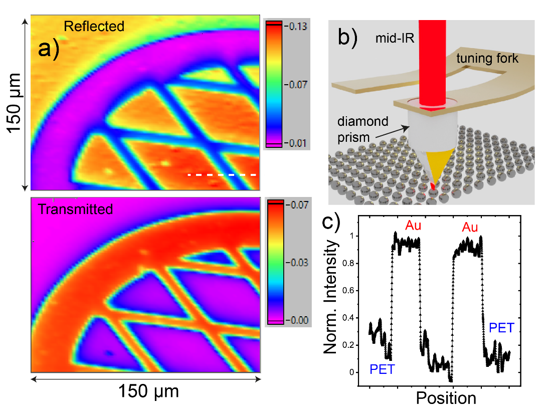
Figure: a) Intensity mappings of simultaneously measured reflected (top panel) and transmitted (bottom panel) signals when a focused 9 μm light beam is moved along the sample (patterned 120 nm thick Au deposited on polyethylene terephthalate (PET)) surface. Here, the focused light spot size is ~30 μm. b) Illustration shows the focusing of mid-IR light through campanile Au/diamond/Au prism connected to advanced electromechanical LiNbO3 tuning fork. c) Normalized intensity measured via reflection when the campanile probe is moved along the dashed line shown in top panel of (a). The observed change in intensity between the Au and PET regions is ~1.5 %.
References:
- H. Choo et al., Nature Photonics 6, 838 (2012).
- M. Mrejen et al., Nature Commun. 6, 7565 (2015).
This work is supported by the UKRI HiWiN research grant and FELIX-Nijmegen, Radboud University.
- References
- H. Choo et al., Nature Photonics 6, 838 (2012).
- M. Mrejen et al., Nature Communications 6, 7565 (2015).