In situ (S)TEM Study of Thermal Reduction Synthesis Pathway for Sulfur Containing Titanium MAX Phase to MXene Phase
- Abstract number
- 260
- Presentation Form
- Poster & Flash Talk
- DOI
- 10.22443/rms.mmc2023.260
- Corresponding Email
- [email protected]
- Session
- Poster Session One
- Authors
- Mr Joseph John Burman (2, 3), Dr Mounib Bahri (2, 3), Mr Ioannis Siachos (2, 3), Prof. Volker Presser (1), Prof. B. Layla Mehdi (2, 3)
- Affiliations
-
1. INM – Leibniz-Institute für Neue Materialien gGmbH
2. University of Liverpool
3. Albert Crewe Centre, University of Liverpool
- Keywords
MXene, MAX phase, Electron Microscopty, Transmission Electron Microscopy, Scanning Transmission Electron Microscopy, In situ, ex situ, In situ techniques, In situ gas, Ins itu STEM, in Operando in Operando STEM, High resolution TEM
- Abstract text
Two-dimensional (2D) nanomaterials with differing compositions and morphologies have long been of interest to a wide range of scientific communities. MXenes, a relatively new material of this class, first synthesised in 2011 display a large range of properties associated with 2D nanomaterials. Notable attributes include high electron density at the Fermi level, ferro- and anti-ferromagnetic capabilities, low resistance, mechanical strength, functional redox potentials, and intercalation capabilities. This range of properties lends them to be applicable to the contemporary topic of energy storage with promising research into applications in pseudo-capacitors and Li-ion batteries. [1]
MXene structures are a group of transition metal nitrides, carbides and carbonitrides with the empirical formula Mn+1XnTx (n = 1, 2, or 3) where M, X and T are: early d-block transition metals, either (and/or) C or N atoms and surface termination groups (commonly F, OH and/or O) respectively, where x is the number of terminations. Owing their name to MAX phase structures (in this case Ti2SC) which are precursor materials possessing a hexagonal layered ternary structures with an “A” component accounting for an sp element in group 13 or 14. The “A” component is etched away leaving the MXene. In this work sulfur the “A” component is etched, resulting in Ti2C a mono-transition metal and face-centred cubic layered MXene. [1, 2]
The synthesis pathways for MXene materials have been heavily researched since their conception. Initial methods utilised aqueous HF enchant for the removal the “A” component, followed by exfoliation. [1, 3] Since then, efforts have been made to move to fluorine-free synthesis such as electrochemical and thermal reduction to provide a more economically and ecologically scalable synthesis. [4]
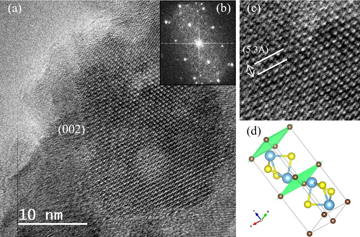
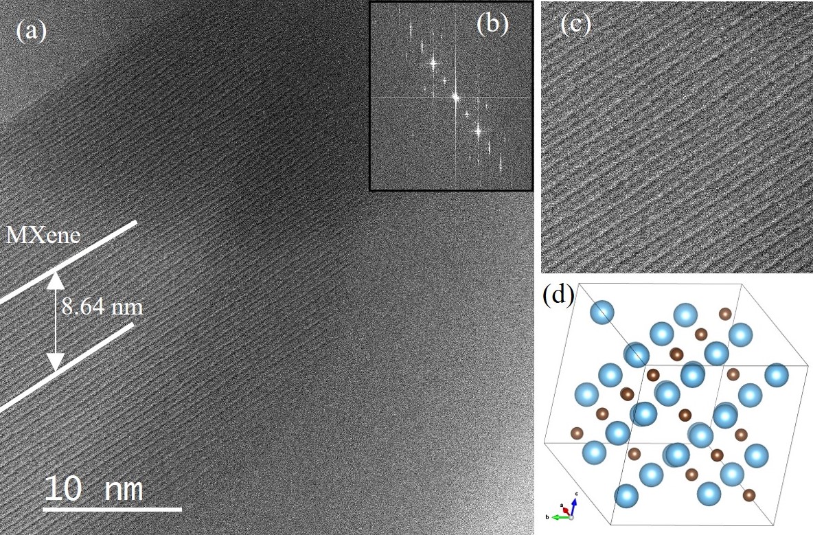
This work endeavours to investigate thermal reduction whilst under in operando conditions utilising both in situ and ex situ approaches to view the synthesis in real time using in situ gas scanning transmission electron microscopy (STEM) in conjunction with residual gas analysis by mass spectroscopy; and post-mortem (ex situ) examinations by transmission electron microscopy (TEM) and energy dispersive x-ray spectroscopy (EDX). Efforts so far have resulted in HR-TEM imaging of the MAX phase Ti2SC viewed down the (002) plane, as seen in Figure 1.(a) and bright field (S)TEM images of in situ gas experiments at elevated temperatures showing the 2D laminated structure of the Ti2C MXene as seen in Figure 2.(a). Post-mortem EDX of MAX phase samples treated under H2/Ar conditions at 700 °C for 1, 6 and 10 hours have indicated a reduction of sulfur atomic percentage over time as expected with the transition from MAX phase to MXene.
Thus far, research shows the differences in the structures between the MAX phase and MXene phase produced by thermal reduction highlighted by (S)TEM microscopy characterisation as seen in Figure 1(a) and 2(a). [5] Validity has been further corroborated due to Figure 2(a) being the result of an in situ study highlighting thermal reduction as a viable synthesis method for greener MXene production on the nano-scale. Further development to this work aims to reveal details of the phase and structural changes in the transition from MAX phase to MXene and identify any possible transitional states that occur between the extrema of the figures shown.
Figure 1: (a) High resolution TEM image of MAX phase as viewed down the (002) plane. (b) Corresponding FFT of image (a). (c) A reduced area showing the atomic resolution of the 5.3 Å d-spacing of the (002) plane of Ti2SC. (d) Depicts the MAX phase unit cell of Ti2SC with the green planes indicating the (002) and repeat. [5]
Figure 2: (a) In situ bright field STEM image under Ar/H2 at elevated temperature showing a measured spacing corresponding to 18 layers. (b) The corresponding FFT of the STEM image. (c) A reduced area of image (a) showing the result of synthesis of the 2D MXene layered structure. (d) Depicts the cubic Ti2C MXene unit cell oriented to match the layered structure shown in (c). [5]
- References
[1] M. Naguib, M. Kurtoglu, V. Presser, et al., Adv. Mater. Vol. 23 (2011) Pages 4248-4253. doi:10.1002/adma.201102306
[2] J.-C. Lei, X. Zhang and Z. Zhou, Front. Phys. Vol. 10 (2015) Pages 276-286. doi: 10.1007/s11467-015-0493-x
[3]O. Mashtalir, M. Naguib, V. N. Mochalin, et al., Nat Commun Vol. 4 (2013) Pages 1716. doi:10.1038/ncomms2664
[4] N. H. Solangi, R. R. Karri, S. A. Mazari, et al., Coord. Chem. Rev. Vol. 477 (2023) Pages 214965. doi: 10.1016/j.ccr.2022.214965
[5] This work has been conducted at the Albert Crewe Centre for Electron Microscopy at the University of Liverpool, UK.