Towards a 100,000 frames per second direct detector for 4D STEM
- Abstract number
- 93
- Presentation Form
- Submitted Talk
- DOI
- 10.22443/rms.mmc2021.93
- Corresponding Email
- [email protected]
- Session
- Stream 2: EMAG - Instrumentation Development (incl Detector technology)
- Authors
- Dr Barnaby Levin (1), Debaditya Chatterjee (2), Jingrui Wei (2), Dr Alexander Kvit (2), Dr Benjamin Bammes (1), Dr Robert Bilhorn (1), Prof Paul Voyles (1)
- Affiliations
-
1. Direct Electron LP
2. University of Wisconsin Madison
- Keywords
Direct Detection
4D STEM
Ultrafast
- Abstract text
Four-dimensional scanning transmission electron microscopy (4D STEM) is a powerful technique that can offer high contrast imaging at low dose [1], drastically improved spatial resolution [2], and a wide range of other unique imaging signals [3]. One current limitation of 4D STEM is its relatively slow speed of acquisition. Typical 4D STEM detectors offer frame rates on the order of 103-104 fps, about 2 orders of magnitude slower than the speed of conventional STEM imaging, which is of order 105-106 probe positions per second.
Here, we report the development of a new, ultrafast monolithic active pixel sensor (MAPS) based direct detector named Celeritas, which has been specifically designed for 4D STEM. The first Celeritas detector is installed on a Cs-corrected Titan STEM at the University of Wisconsin-Madison Nanoscale Imaging and Analysis Center, a user facility open to scientists from around the world, where initial testing has taken place. The physical sensor is 1024×1024 pixels, with 15 mm pixel size. At full frame readout, the detector exceeds 2,100 frames per second (fps). Sub-area readout enables higher frame rates, up to an anticipated maximum exceeding 105 fps – comparable to conventional STEM imaging speeds – at an area of 256×64 pixels. Sub-area readout can be configured in software, allowing users to trade pixel count for speed to suit different applications. Additional features incorporated into Celeritas include rolling shutter and global shutter readout modes, and on-chip correlated double sampling (CDS), a method that subtracts reset noise from each pixel in each frame to increase the signal to noise ratio (SNR) of the detector.
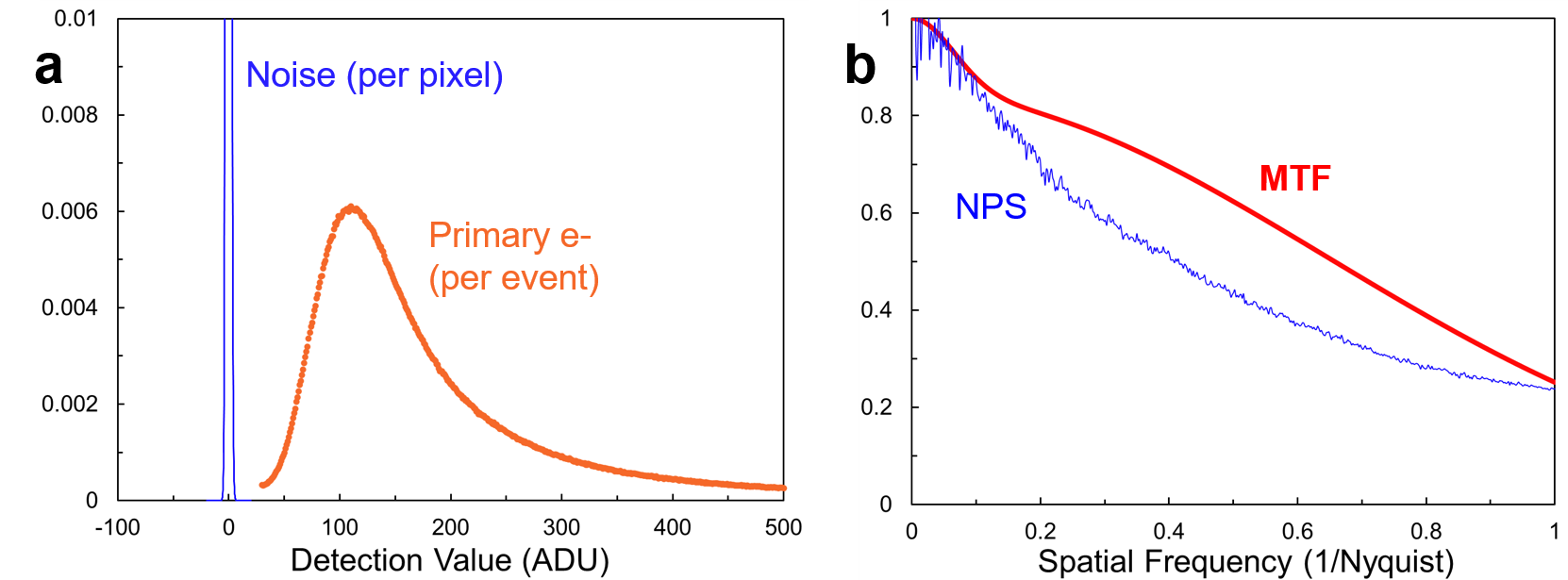
Fig. 1. summarizes basic detector performance. Fig. 1a shows that the mean and most probable intensities of single 200 keV primary electrons are 266 analog-digital units (ADU) and 110 ADU respectively in rolling shutter mode with on-chip CDS enabled. In the absence of incident electrons, the root-mean-squared pixel value is just 1.32 ADU. This corresponds to a mean SNR of ~200:1 and most probable SNR of ~80:1 for single electrons. The mean detection event size for 200 kV electrons has been measured as 2.5 pixels which yields the modulation transfer function (MTF) in Fig. 1b. MTF exceeds 0.6 at half Nyquist when the camera is operated in integrating mode. As with other MAPS detectors, electron counting can be applied to sparse images to normalize the signal of individual detected electrons and further increase MTF. Fig. 2 shows initial 4D STEM data from a [100] SrTiO3 sample, acquired at 1,800 fps with full frame readout in high gain mode, with a 39 pA probe current, 23.4 mrad convergence semi-angle, and 34 mrad maximum collection angle. Fig. 2a is a typical single CBED pattern from the dataset. Fig. 2b is a synthetic bright-field image, showing the Sr and Ti sublattices of the SrTiO3. Fig. 2c is the 90-degree rotation symmetry STEM image [4], showing sharp maxima at atomic column positions.
For some 4D STEM applications, it is important to simultaneously image the intense central beam and sparse high angle scattering of a convergent beam electron diffraction pattern. This can be challenging for typical MAPS based direct detectors, which saturate between 100 and 101 e-/pixel per frame. To address this challenge, Celeritas can be operated in different gain modes. In high-gain mode, saturation occurs at 10 e-/pixel per frame, whilst in low-gain mode the SNR for single electron events is reduced, but the saturation level exceeds 130 e-/pixel per frame. At 105 fps, this is equivalent to ~2 pA/pixel, which is sufficient to measure the central beam without saturation, even at short camera lengths. A high dynamic range (HDR) mode, in which the same exposure is read in both low- and high-gain modes at a cost of a 2× reduction in maximum framerate allows high SNR for weak signals and high saturation for strong signals. This can be combined with a patent-pending HDR counting algorithm that applies electron counting in sparse, dark regions and integration in bright regions of each frame [5], to further improve SNR.
With the high speeds enabled by Celeritas, a number of potential experiments become more practical. One example is time-resolved 4D STEM, where the time between entire 4D STEM acquisitions could be as little as 0.2 s for 128×128 probe position datasets. Furthermore, high speeds can enable 4D STEM to outrun many instrument instabilities, so techniques developed to improve SNR and reduce spatial distortions in conventional STEM, like non-rigid registration [6] and RevSTEM [7] could be applied, pointing toward high precision 4D STEM. Intended future applications of Celeritas include electron correlation microscopy [8] atomic dynamics measurements in supercooled liquids and ptychographic movies of defects in materials at elevated temperature.
Direct Electron acknowledges support from the US Department of Energy, Office of Science, grant DE-SC0018493. Development of the ultrafast Celeritas detector was also supported by the Wisconsin MRSEC (DMR-1720415).
Figure 1: (a) Histograms of background noise per pixel per frame in the absence of incident electrons and the total signal deposited in the detector by incident 200 kV primary electrons in rolling shutter high-gain mode at ~1800 fps. (b) MTF and noise power spectrum (NPS) for 200 kV electrons.
Figure 2: 4D STEM data acquired with Celeritas from a SrTiO3 sample at ~1,800 fps, full-frame. (a) A typical single frame CBED pattern. (b) A synthetic bright-field STEM image. (c) A 90-degree symmetry STEM image. The color scales in (a) and (b) are in detector counts. The color scale in (c) is the value of the symmetry coefficient.
- References
[1] C. Ophus, Microsc. Microanal. 25, 563 (2019).
[2] L. Zhou, et. al. Nat. Commun. 11, 2773 (2020) .
[3] Y. Jiang, et. al., Nature 559, 343 (2018).
[4] M. Krajnak, J. Etheridge, Proc. Natl. Acad. Sci. 117, 27805 (2020).
[5] B.D.A. Levin, et. al. , Microsc. Anal. 34(1), 20 (2020).
[6] A.B. Yankovich, et. al. Nat. Commun. 5, 4155 (2014).
[7] X. Sang, J. M. Lebeau, Ultramicroscopy 138, 28 (2014).
[8] L. He, et. al., Microsc. Microanal. 21, 1026 (2015).


