Tools for spatial analysis to understand the immunopathology associated with infectious disease.
- Abstract number
- 302
- Presentation Form
- Submitted Talk
- Corresponding Email
- [email protected]
- Session
- Stream 5: High-plex Cytometry
- Authors
- Dr Helen Ashwin (2), Dr Grant Calder (1), Dr Sally James (1), Dr Lesley Gilbert (1), Dr Karen Hogg (1), Dr Mohamed Osman (2), Prof Paul Kaye (2)
- Affiliations
-
1. Biosciences Technology Facility, Department of Biology, University of York
2. York Biomedical Research Institute, Hull York Medical School, University of York
- Keywords
Spatial, Image analysis, StrataQuest
- Abstract text
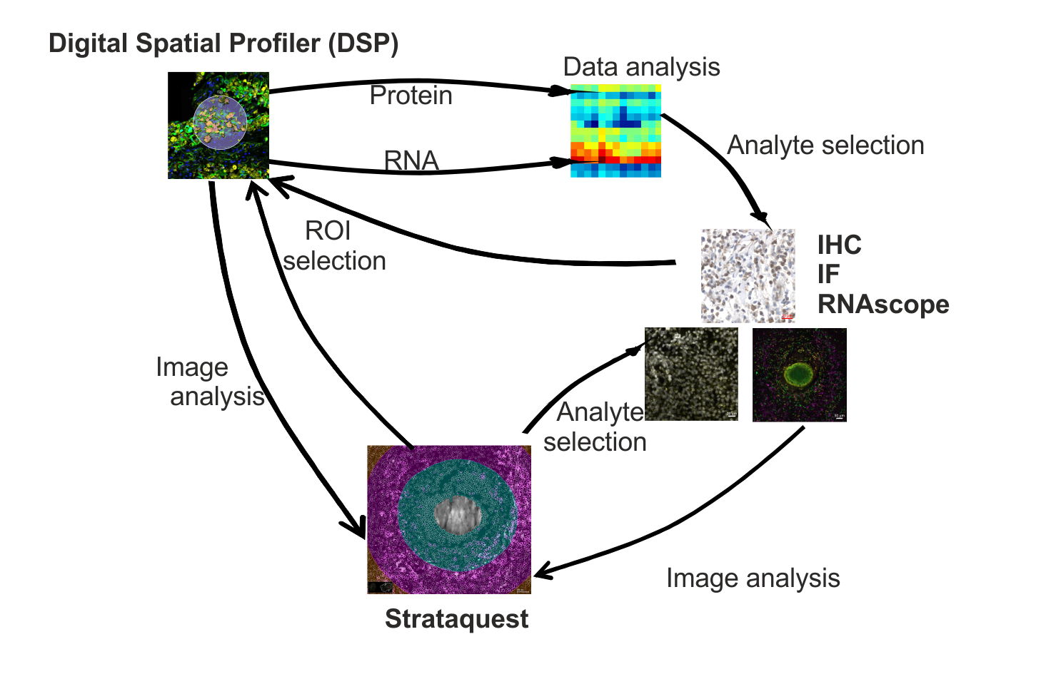
We used image analysis software StrataQuest (TissueGnostics) to automatically quantify a range of features within histological sections imaged on a slide scanner (Axio Scan.Z1, Zeiss). These sections have either been stained using immunohistochemistry (IHC), immunofluorescence (IF) or RNAscope® probes alongside tinctorial stains (e.g. trichrome, haematoxylin and eosin).
Using StrataQuest we have context-based quantitative analysis for cell number, density and type and the spatial relationship to tissue structures, e.g granulomas and epidermis. These analyses pipelines help guide and support conducting highly multiplexed spatial omics analysis by nanoString GeoMx® Digital Spatial Profiler (DSP) (e.g.ROI selection, target validation, whole tissue profiling).
We will discuss these approaches in the context of ongoing work on the pathology associated with COVID-19 and neglected diseases of parasitic (leishmaniasis) as well as fungal and bacterial origin (mycetoma).
Figure 1: Workflow linking StrataQuest image analysis, digital spatial profiling and histology.