Ptychography on dynamically scattering samples
- Abstract number
- 240
- Presentation Form
- Submitted Talk
- DOI
- 10.22443/rms.mmc2021.240
- Corresponding Email
- [email protected]
- Session
- Stream 1: EMAG - 4D-STEM
- Authors
- Dr Laura Clark (4), Dr Gerardo Martinez (1), Dr Colum O'Leary (3), Dr Tim Petersen (2), Dr Scott Findlay (2), Prof Peter Nellist (5)
- Affiliations
-
1. IMEC
2. Monash University
3. University of California
4. University of Leeds
5. University of Oxford
- Keywords
4D-STEM, DPC, ptychography, dynamic scattering, phase objects
- Abstract text
Phase imaging of specimens in the STEM is becoming increasingly popular with the growing access to pixelated electron detectors – with relevant imaging techniques spanning across differential phase contrast (DPC) based methods and ptychographic techniques. Direct (non-iterative) electron ptychography in particular is rapid [1], dose-efficient [2] and can be run live at the microscope [3]. However, much of the theory is built upon the (weak) phase object approximation, and the projection approximation [4]. These criteria are strictly fulfilled by a limited subset of the samples we may wish to study. This naturally raises the question – how reliable are these methods when applied to the much wider classes of thicker specimens?
In this work, we present results from an investigation into applications of STEM phase imaging on a thickness-series of simulated data. Contrast from Wigner distribution deconvolution (WDD) and single sideband (SSB) ptychography remains on-column for dynamically scattering samples, beyond thicknesses where other methods lose column-resolution. This is confirmed with reference to experimental data. Furthermore, the ability to post-correct aberrations in experimental data allows for clear atomic-resolution images even when the collection conditions are not ideal –such as when collecting data on beam-sensitive samples. Following from this, we indicate the optimum focal conditions for collecting 4D-STEM phase imaging experimental data.
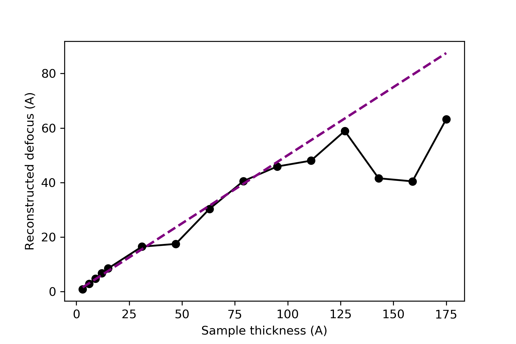
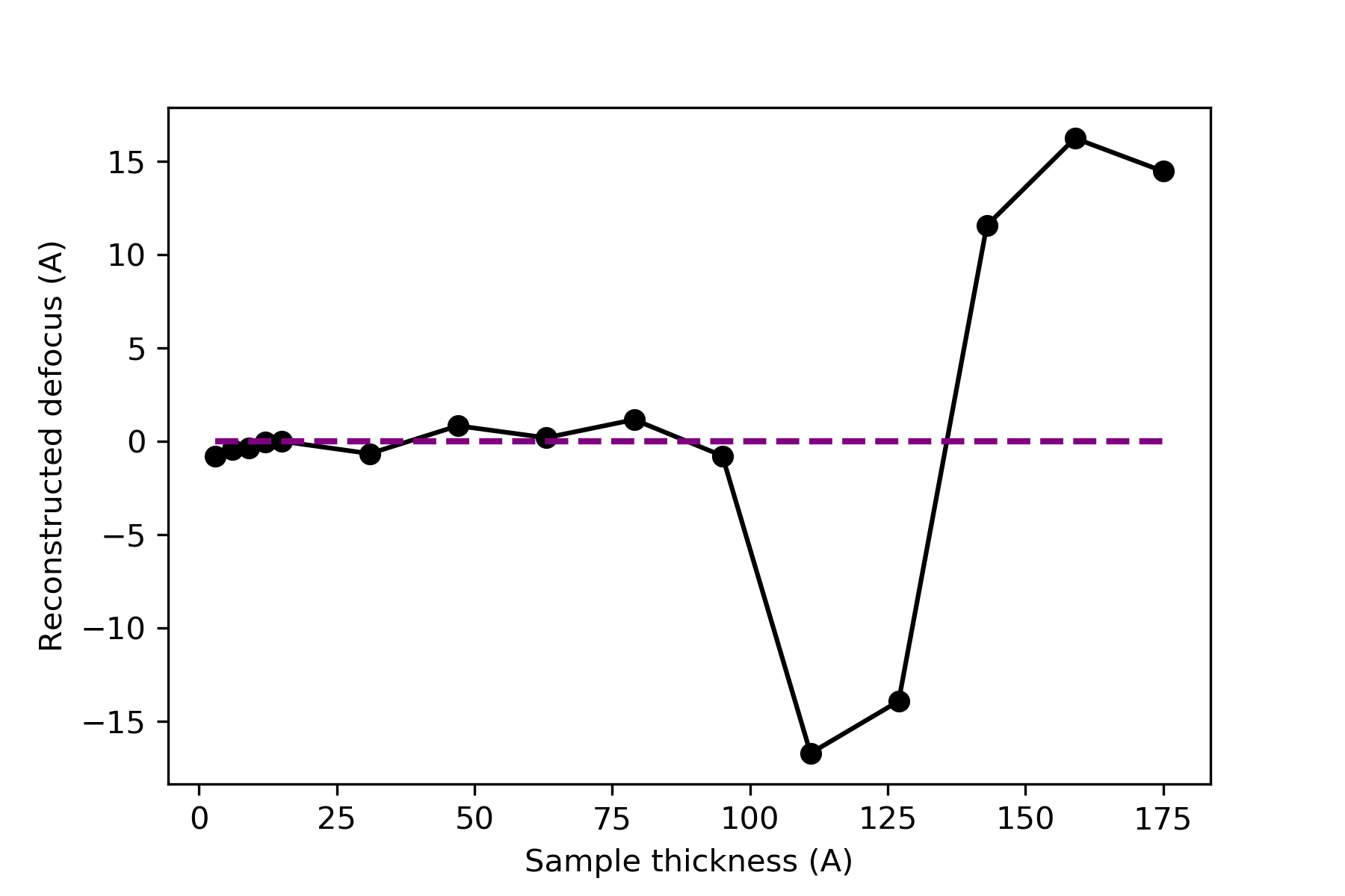
Simulations through a thickness series of GaN allow a comparison of conventional BF and DF images with (integrated) DPC, SSB and WDD ptychography. Analysis of the 4D Fourier transformed data of these simulations reveal aberrations in the dataset, minimised at a mid-sample focal condition as indicated in the figures below.
Figure 1: Reconstructed defocus analysis from a thickness series of GaN simulations (solid black line). Indication of defocus equal to thickness/2, purple dashed line. Data from the top surface focal condition, WDD analysis.
Figure 2: Reconstructed defocus analysis from a thickness series of GaN simulations (solid black line). Indication of defocus equal to zero, purple dashed line. Data from the midplane focal condition, WDD analysis.
- References
[1] CM O’Leary et al. "Phase reconstruction using fast binary 4D STEM data." Applied Physics Letters 116.12 (2020): 124101.
[2] TJ Pennycook et al. "High dose efficiency atomic resolution imaging via electron ptychography." Ultramicroscopy 196 (2019): 131-135.
[3] K Muller-Caspary, “Momentum-resolved STEM: An interdisciplinary methodological platform for materials and soft matter characterization.” RMS Virtual Microscopy Characterisation of Organic-Inorganic Interfaces, 12th March 2021.
[4] M Vulović et al. "When to use the projection assumption and the weak-phase object approximation in phase contrast cryo-EM." Ultramicroscopy 136 (2014): 61-66.


