Introducing the M4 (MultiModal Modular Microscopy) 3D Printed Microscope System for Imaging Macrophage-Pathogen Interactions
- Abstract number
- 283
- Presentation Form
- Submitted Talk
- DOI
- 10.22443/rms.mmc2021.283
- Corresponding Email
- [email protected]
- Session
- Stream 5: Up Close with the Enemy: Imaging Pathogen-host Dynamics
- Authors
- Miss Gemma Cairns (2, 1), Prof David Dockrell (1), Dr Brian Patton (2)
- Affiliations
-
1. University of Edinburgh
2. University of Strathclyde
- Keywords
Macrophages, Streptococcus pneumoniae, Host-Pathogen Interaction, 3D Printing, Open Source Microscopy, Microscope Development, Fluorescence
- Abstract text
Mitochondrial reactive oxygen species (mROS) have recently emerged as critical microbicidal factors employed by macrophages1,2. Our long-term aim is to quantify the role of mROS in the clearance of internalised extracellular bacteria in alveolar macrophages, particularly Streptococcus pneumoniae, the most common cause of the inflammatory condition pneumonia2. Recently, colocalisation has been observed between mROS, generated in response to S. pneumoniae, phagolysosomes and the bacteria itself2.
Advances in microscopy and image analysis offer new opportunities to quantify these interactions to allow future examination of our ability to pharmacologically enhance host responses against antimicrobial resistant bacteria. However, there are numerous challenges posed when trying to image mROS in macrophages, particularly when also trying to correlate mROS location and dynamics with specific host responses.
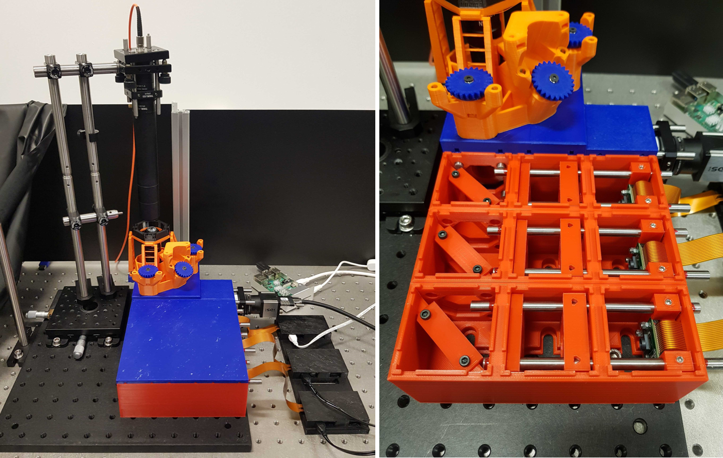
To address some of these challenges, we have developed the M4: MultiModal Modular Microscopy system, a fully open-source 3D-printable optics system which allows users to design and build different setups for different imaging modalities. Advanced microscopy hardware, for techniques such as live cell and super-resolution microscopy, which are key for investigating host-pathogen interactions, can be costly and often inaccessible to research groups. Frugal innovation will therefore allow an increased availability of advanced microscopy technologies, particularly when combined with computational microscopy techniques.
For colocalisation studies in cells, for example between mitochondria and S. pneumoniae, it is necessary to use multichannel fluorescence microscopy. Here, we present the M4 system in combination with the OpenFlexure3 stage, as seen in the figure below, and alignment/calibration tools based on ray transfer matrix analysis for building a multichannel imaging system. We also present initial imaging data of macrophages using a multichannel fluorescence microscope built with the M4 system. In future we hope to employ the M4 system within a cell culture incubator for live cell imaging of macrophage-pathogen interactions.
- References
- West, A. P. et al. TLR signalling augments macrophage bactericidal activity through mitochondrial ROS. Nature 472, 476-480 (2011).
- Bewley, M. A. et al. Impaired Mitochondrial Microbicidal Responses in Chronic Obstructive Pulmonary Disease Macrophages. Am. J. Respir. Crit. Care Med. 196, 845-855 (2017).
- Sharkey, J. P. et al. A one-piece 3D printed flexure translation stage for open-source microscopy. Rev. Sci. Instrum. 87, 25104 (2016).