Automated In-Situ Spectrum Imaging with Synchronized Stimulus Control
- Abstract number
- 224
- Presentation Form
- Poster
- Corresponding Email
- [email protected]
- Session
- Poster Session 3
- Authors
- Dr Liam Spillane (1), Dr Benjamin Miller (1), Dr Bernhard Schaffer (1), Dr Paul Thomas (1), Dr Ray Twesten (1)
- Affiliations
-
1. Gatan Inc.
- Keywords
STEM, EELS, EDS, CBED, 4DSTEM, in-situ, heating, automation, multimodal, DMScript, Python, Scanning Transmission Electron Microscopy, Electron Energy-Loss Spectroscopy, Diffraction, Convergent Beam Electron Diffraction, Catalysis, Metal Oxide, Electron Counting, Counting EELS,
- Abstract text
The scanning transmission electron microscope (STEM) is a well-established analytical tool for characterizing material structure-property relationships due to the large number of signals result from the beam-specimen interaction, combined with the fact that many can be acquired simultaneously at high spatial resolution.
Spectrum imaging (SI), a modality enabling the acquisition of spatially resolved analytical signals, is particular powerful due its capability to enable direct correlation between imaging and analytical data. Three primary types of STEM-SI have been widely adopted. Diffraction spectrum imaging (4DSTEM, CBED-SI) gives morphological and crystallographic information. Energy dispersive (x-ray) spectroscopy (EDS) SI, and electron energy-loss spectroscopy (EELS) SI give compositional information. EELS can also be used to probe local electronic structure, allowing local determination of: bonding states, chemistry, phonon and/or electronic band structure. For in-situ STEM-SI analysis, multiple SI passes must be acquired, making the ability to record data at high speed and high dose efficiency particularly important [1]. In order to avoid missing important transformations during an experiment, high in-situ stimulus resolution is also of fundamental importantance [1].
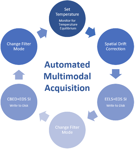
In this work, we use a CMOS based transmission electron counting detector fitted into an optimized post-column energy filter (K3 Continuum-IS) to demonstrate recent advances in synchronized in-situ spectrum imaging acquisition. These advances allow fully automated acquisition of the CBED and EDS signals at a given in-situ condition, in addition to the EELS signal, maximizing the information that can be extracted from a single specimen and target region of interest Additionally, datasets are now streamed/written directly to disk rather than held memory in over the duration of the experiment, following the workflow steps shown in figure 1. The combined effect of these advances maximizes the information that can be extracted from a single specimen, even for irreversible transformations as all data can be acquired in a single experiment, in addition to maximizing the stimulus resolution at which the data can be captured.
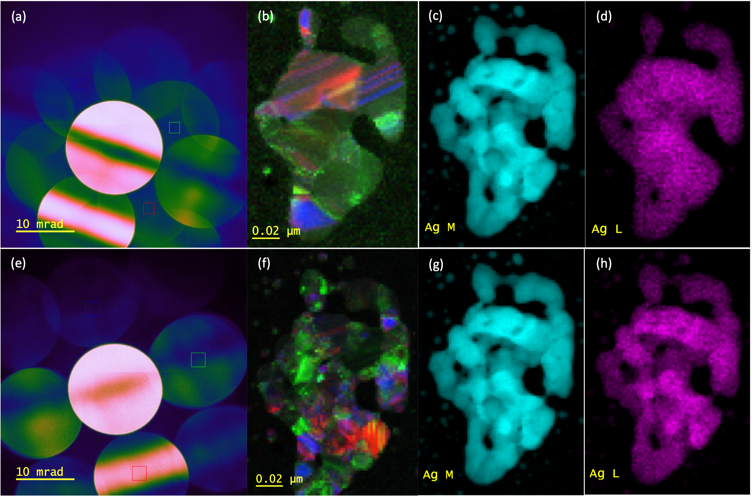
Metal oxide systems have been used as model materials systems. Example dual-EELSTM, energy-filtered diffraction and EDS multipass spectrum image results acquired from silver (II) oxide over temperature range 25 – 600°C are shown in figure 2. Temperature control was implemented as per the flow diagram show in figure 1. In this presentation we will expand upon these findings to highlight the importance and impact of automation and synchronization to in-situ STEM analysis.
Figure 1. Flow diagram showing data acquisition and holder control steps in the multimodal multiple pass spectrum imaging method.
Figure 2. Multimodal SI data acquired from silver(II) oxide powder at 600°C: (a) energy filtered CBED pattern showing virtual aperture positions for (b) orientation map. (c) EELS Ag M-edge map, and (d) EDS Ag L-peak map. Equivalent data for 25°C is also shown (e-h).
- References
[1] L. Spillane et al., Microsc. Microanal. 26 (Suppl 2), (2020)