Towards a better understanding of corrosion processes in zirconium alloys through correlated serial sectioning using laser PFIB
- Abstract number
- 329
- Presentation Form
- Contributed Talk
- DOI
- 10.22443/rms.mmc2023.329
- Corresponding Email
- [email protected]
- Session
- EMAG - 3D & Tomographic Electron Microscopy
- Authors
- Dr Alistair Garner (1), Dr Sam Armson (1), Dr Jack Donoghue (2), Dr Aidan Cole-Baker (1)
- Affiliations
-
1. Jacobs
2. University of Manchester
- Keywords
3DEBSD, corrosion, laser PFIB, zirconium, nuclear materials
- Abstract text
A novel methodology has been developed to investigate the effect of substrate microstructure on oxide growth mechanisms in Zr alloys, used as cladding material in nuclear reactors. The corrosion of Zr alloys has been the subject of an intensive research effort over the past ~50 years, and corrosion of Zr alloys is currently one of the main factors limiting the lifetime of reactor fuel assemblies. Improved understanding of corrosion process can therefore lead to the development of more corrosion resistant alloys, more efficient power generation, and reduced waste levels.
The technique involves initial mechanical preparation of an oxidised block of a hydrided commercial Zr alloy to produce a 200 µm thick sheet. The fs-laser of the laser-PFIB is then used to machine away large regions of the surrounding material away in the order of minutes to isolate a block ~300 µm across to allow the escape of both milled material and EBSD patterns. The block is then serial sectioned with high positional accuracy using the Xe ion beam, with the oxide on the bottom face, to reduce milling artefacts encountered when the beam hits the oxide first.
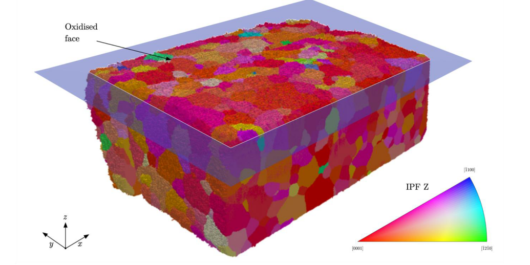
Figure 1: 3D-EBSD volume showing Zr substrate microstructure. Grains are shown in inverse pole figure colouring parallel to sample normal direction (IPF-Z) according to legend.
An EBSD map and a corresponding high resolution secondary electron image are collected on every second slice. These datasets are used to create a 3D reconstruction of both the crystallographic nature of the zirconium substrate (Figure 1), and the morphological nature of the zirconium oxide film that has formed during corrosion (Figure 2). The 3D EBSD dataset contains information on the overall crystallographic grain orientations, internal and intergranular misorientations as well as the morphology (and in some cases orientation) of hydrides present in the material prior to corrosion. These features can then be correlated to the oxide morphology in three dimensions, both in terms of local oxide thickness and internal oxide cracking.
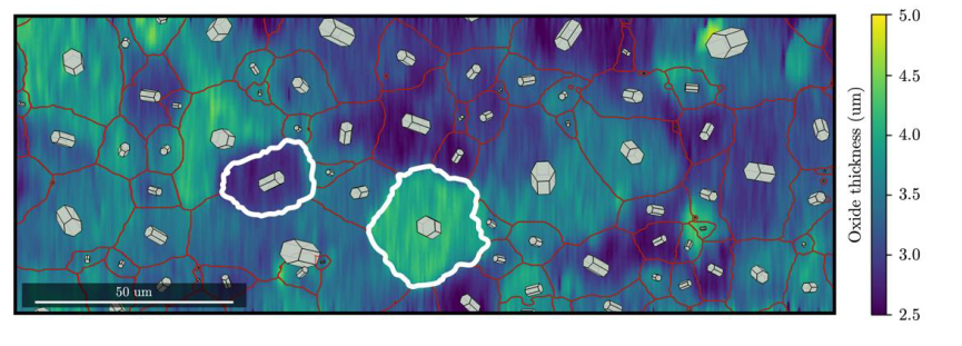
Figure 2: Oxide thickness map overlaid with metal grain boundaries and unit cell schematics showing effect of substrate orientation on resulting oxide thickness.
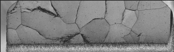
It was observed that the local oxide thickness is highly sensitive to the underlying substrate grain orientation, and the degree of oxide protectiveness linked to the deviation of the c-axis away from the oxide growth direction (Figure 2). In addition, the presence of specific grain boundaries has been correlated to local differences in oxide morphology and internal cracking. The presence of hydrides in the metal has also been observed to disturb the usual oxide growth processes. The analysis has allowed for a high resolution 3D reconstruction of hydride morphology which has shown the complex nature of these hydride clusters in unprecedented detail which can then be correlated to the Zr grain orientations from which they formed (Figure 3).
Figure 3: Band contrast map showing complex hydride morphology in single slice
These types of studies are usually performed using TEM, which typically lack a statistically significant number of observations, or by plan view EBSD which makes it difficult to directly relate the substrate and oxide orientations, and does not allow for imaging of defects within the oxide. The correlative approach developed here allows for high resolution oxide imaging to be combined with large scale 3D imaging over a statistically significant number of substrate grains. The results have been used to provide new insight into oxide growth mechanisms and in particular the dominant effects of substrate microstructure on resulting oxide formation. It is also believed that the technique can be applied to advance understanding in a number of other corrosion and thin film applications across the materials science community.