Advanced Cryogenic Focussed Ion Beam Sample Preparation: Beyond Electron Microscopy
- Abstract number
- 441
- Presentation Form
- Poster & Flash Talk
- DOI
- 10.22443/rms.mmc2023.441
- Corresponding Email
- [email protected]
- Session
- Poster Session Two
- Authors
- Dr Chris Parmenter (2), Ms Haneen Packeer Ally (2), Dr Julie Watts (2), Dr Ken Fahy (1), Dr Lucian Kaack (3), Dr Steven Jansen (3), Dr Lisa White (2)
- Affiliations
-
1. SiriusXT
2. University of Nottingham
3. University of Ulm
- Keywords
Cryo-FIB, Cryo-Lift-out, Soft Matter, Tomography
- Abstract text
Summary
We present recent efforts to use Cryogenic Focused Ion Beam Scanning Electron Microscopes (cryo-FIB-SEM) as a preparation tool for cryogenic analysis beyond the FIB itself, embracing emerging themes in multi-length-scale analysis and correlative microscopy. Cryogenic FIB preparation combined with lift-out using an in situ cryogenically cooled micromanipulator is demonstrated as a preparation tool for cryo-Transmission EM (cryo-TEM), soft X-ray (sXT), and X-ray Nanotomography (XNT) systems and can be further expanded to Atom Probe Tomography preparation (APT)
Introduction
Focused Ion Beam Scanning Electron Microscopes (FIB-SEM) used under Cryogenic (cryo) conditions have been used as a preparation tool for soft-matter samples since the mid-2000’s, with a rapid expansion of their use in the past five years, particularly in the case of the ‘on-grid’ thinning approach for cryo-electron tomography (cryo-ET) by the biology community. Less well developed is the site-specific FIB preparation (under cryo-conditions) of samples that cannot be grown (or deposited) and thinned on a TEM grid. Such samples can be considered to be bulk samples and may include tissue or larger soft samples. To prepare specific areas for these samples FIB preparation followed by extraction (lift-out) is a route to achieving the specific dimensions and thickness requirements of the TEM prior to transfer. This approach can be expanded to other analytical equipment, subject to meeting their dimensions and requirements and this challenge is explored in this work.
Methods/Materials
This work was performed using Crossbeam 550 FIB-SEM (Carl Zeiss) and Quanta 3D (FEI Company) using a Quorum 3010 cryo-system. The micromanipulator was an Omniprobe 100-cryo (Oxford Instruments). In all cases, samples (yeast and plant material) were frozen by slushy nitrogen, metal mirror (MM) freezing or high-pressure freezing (HPF) before transfer to the cryo-system. Samples were coated in platinum in the preparation chamber and then subsequently by the organometallic precursor of the gas injection system (GIS) of the FIB. FIB preparation was performed at 30kV accelerating voltage and SEM of the samples was performed at 5-15 kV. Gallium FIB milling of the samples was performed at currents of between 5nA (initial larger volume milling) and 50 pA (final polishing of samples). Cryo-condensation of water from the waster GIS was used to attach the sample to the micromanipulator and support substrates.
Results and Discussion
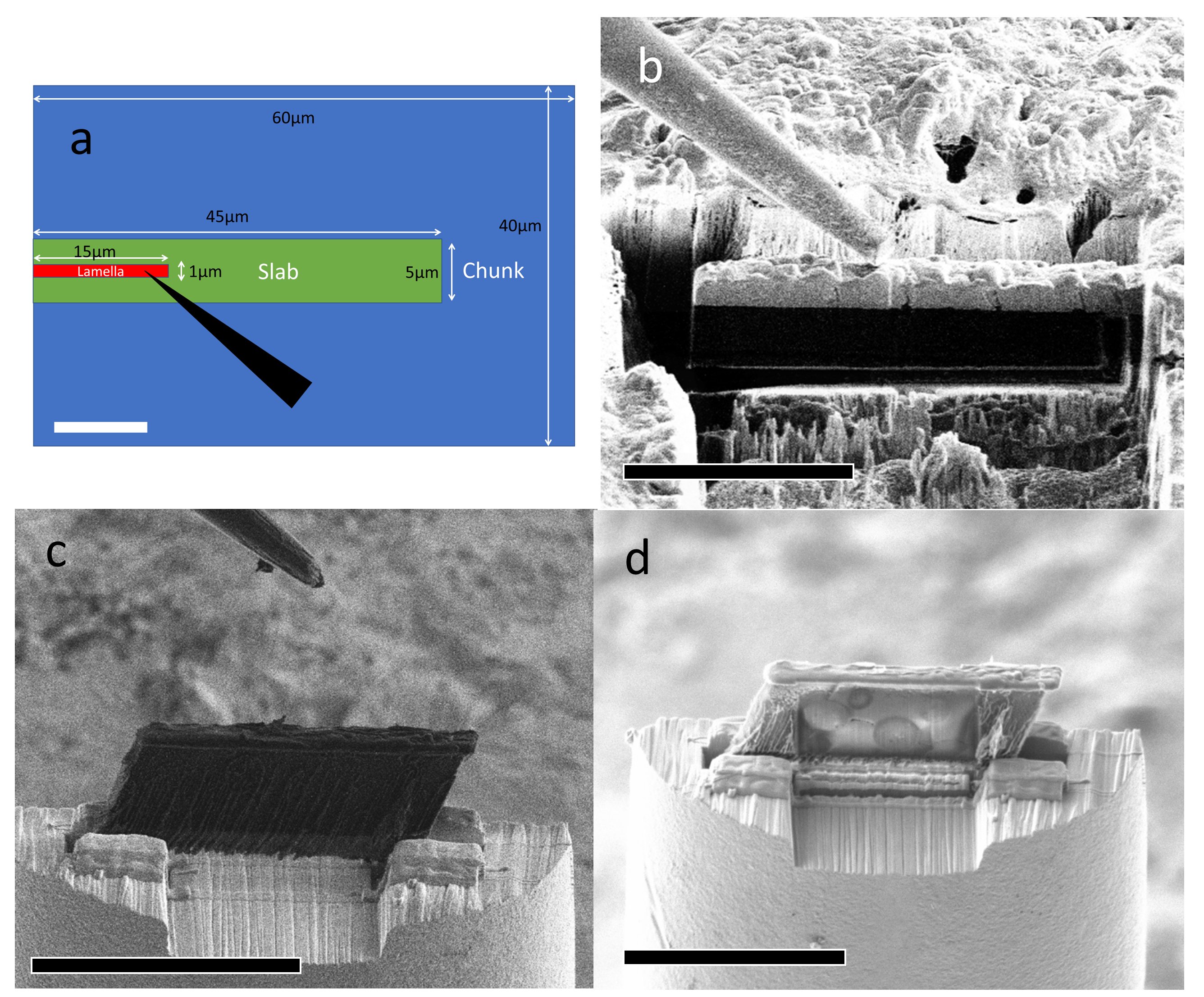
The preparation of cryo-TEM samples was the starting place for this work and built on pre-existing procedures for the preparation of TEM-compliant samples known as lamellae [PARMENTER and NIZAMUDEEN]. Applying experience gained from this we increased the dimensions of the volumes prepared to meet the requirements of the soft-x-ray tomography (sRT) which were approx. 40µm and a thickness of 5µm (fig 1b). The sXT can penetrate a sample up to 10 µm however, we accounted for the sample needing to be tilted in the X-ray beam (+/- 55°). These samples are referred to as ‘slabs’ in contrast to lamellae. These slabs were transferred to specially modified copper TEM support grids with a slot into which the slabs could be inserted (fig 1 c&d).
For compliance with the cryogenic X-ray ptychographic imaging (X-ray nanotomography / XNT) a different sample support (OMNypin) was used to be compatible with the X-ray beamline, which was the eventual destination of the samples. In this case, the dimensions could be considerably thicker due to the higher energies of the X-ray beam and its penetration power. Here samples that we termed ‘chunks’ were produced with dimensions of approx. 60 µm x 40 µm and again extracted using the micromanipulator, after which they were deposited to the OMNYpin and secured with cryo-condensed water vapor. Fig 1a shows the relative dimensions of the various preparation options.
Fig 1. (a) Schematic showing the relative dimensions of the lamella, slab and chunks, the tip of a micromanipulator is included for scale. (b) Ion beam image showing the Omniprobe approaching a slab. (c) Ion beam point of view image following the deposition of a slab onto the support grid. (d) SEM image of a slab with yeast visible. Scale bars (a) 10 µm (b) 20 µm (c&d) 30 µm.
Conclusion
Cryo-FIB combined with cryo-LO has been demonstrated as a viable route to the preparation of a range of soft-matter samples ranging in size from 15µm x 1µm (lamellae for TEM) through 45µm x5µm (slabs for sXT) to 60µm x40 µm (chunks for XNT). This demonstrates that the FIB has an essential role as a preparation tool for forward-looking microscopists keen to understand their samples on a range of instruments with correlative microscopy.
- References
PARMENTER, C.D. and NIZAMUDEEN, Z.A. (2021), Cryo-FIB-lift-out: practically impossible to practical reality. Journal of Microscopy, 281: 157-174. https://doi.org/10.1111/jmi.12953