Multimodal nano-analysis of Cobalt Color Centers in ZnO Nanowires
- Abstract number
- 245
- Presentation Form
- Contributed Talk
- DOI
- 10.22443/rms.mmc2023.245
- Corresponding Email
- [email protected]
- Session
- Correlative and Multimodal X-ray Microscopy
- Authors
- Valentina Bonino (1), Christian T. Plass (2), Maurizio Ritzer (2), Lukas R. Jäger (2), Vicente Rey-Bakaikoa (1), Martin Hafermann (2), Jaime Segura-Ruiz (1), Gema Martínez-Criado (1), Carsten Ronning (2)
- Affiliations
-
1. ESRF − The European Synchrotron Radiation facility
2. Institut für Festkörperphysik, Friedrich-Schiller-Universität Jena
- Keywords
nano-XRF, nano-XEOL, TR-XEOL, synchrotron, nanowires
- Abstract text
The search for single photon sources coupled with the need for compact devices is pushing the research field toward sophisticated fabrication and characterization technologies. Multimodal characterization by means of X-ray fluorescence (XRF), X-ray excited optical luminescence (XEOL) and time-resolved XEOL, excited with a hard X-ray nanoprobe, is here presented for the characterization of color centers in Co-doped ZnO nanowires (NWs) [1].
Color centers in quantum information is a subject of intense research for its application as qubit in quantum computing. In this field, the emission of ZnO can be tuned by transition metals for the fabrication of fast optical sources emitting in a broad visible range. By exploiting this advantage into nanowire (NW) structures one can couple the emission from color centers to the waveguiding and cavity effects of NWs [2]. In these systems, the correlation between the local homogeneity of the chemical and of the optical properties are of importance in the comprehension of the optical dynamics involved and in the optimization of the fabrication parameters.
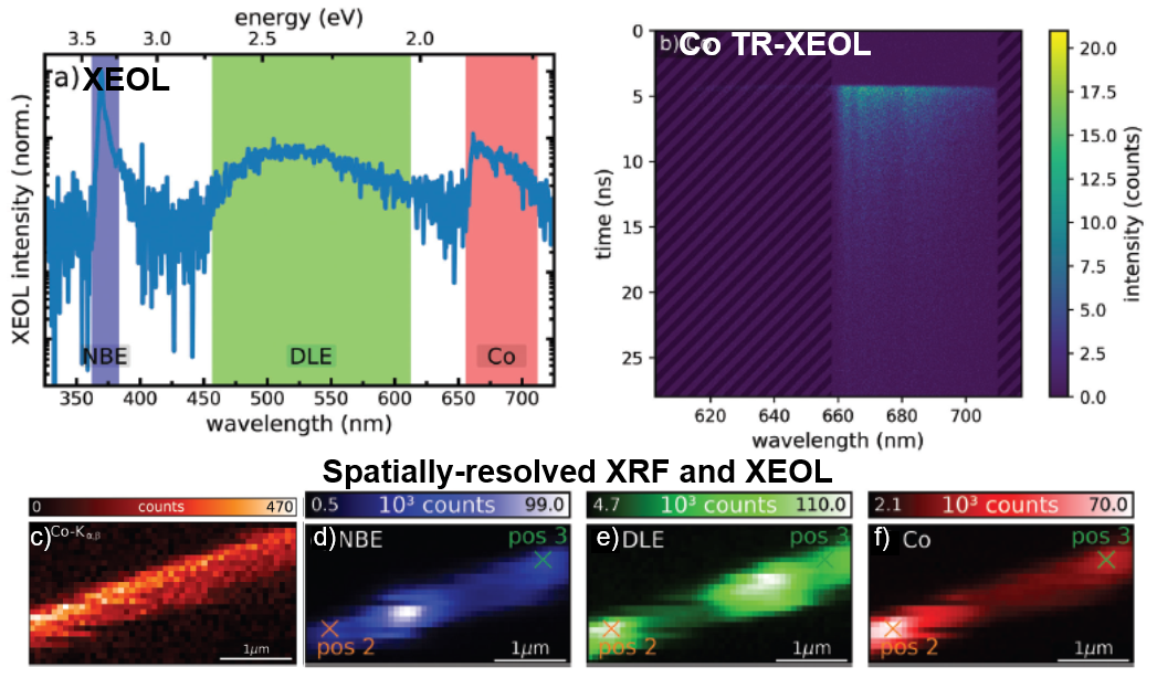
Nanowires of ZnO having a diameter between 200 – 1000 nm were synthesized by vapour-liquid-solid growth mechanism. Cobalt doping was achieved by ion implantation at an energy of 380 keV. Co concentrations of 0.7 at.% were estimated at a depth of about 180 nm from the exposed surface. An annealing at 750 °C was performed to recover the structure from implantation damage. The nanowires were characterized at the nano-analysis beamline ID16B of the European Synchrotron Facility (ESRF), France, after dispersing them on a silicon wafer. A combination of XRF and XEOL measurements were performed by scanning the samples under a nanobeam of 62 × 74 nm2 (V × H) size and a photon flux up to 6 × 10¹² ph/s. The 16-bunch filling mode was used for time-resolved measurements. A streak camera, with an instrument response function of 85 ps, was used to capture in a single image the optical spectrum and its temporal decay in single points of the sample. All the measurements were done at a cryogenic temperature of 10 K.
The Co distribution in the NWs (Figure c) was confined along a depth of about 180 nm, as it was expected from the calculated implantation profiles. The optical emission of the NWs was present in all the probed structures with some variations in the spatial distribution of the intensity. Three optical transitions were detected: the excitonic emission - near band edge (NBE, blue) -, the Co-related emission (red), and the emission from intrinsic defects – deep level emission (DLE, green)- (Figure a). An anticorrelation in their spatial distribution revealed the competitive nature of these transitions (Figure d-f). Moreover, from the comparison of the Co distribution and the optical emissions, it appeared that both structural defects and slightly inhomogeneous Co-implantation profiles were present in the NWs. On the latter point, the tilt of the ion beam, the shadowing effect from adjacent NWs certainly contributed to change the environment of the implanted Co atoms.
The decay time of the dynamics associated to the Co atoms were obtained by fitting the relaxation of the optical signal in time (streak image in Figure b). A double exponential function was used for the fitting, which resulted in a slow decay of 7 ns and a fast decay of 2 ns. While the slow decay was already reported in luminescence measurements [3], the fast decays was observed for the first time. In contrast to the slow decay, which is associated to the transfer of electrons from the conduction band of the ZnO to the 3d energy levels of the Co atoms, the fast decay is most probably reachable only by ionization of core electrons in Co atoms. Such transition would then take place via the direct excitation of the Co atoms by absorption of the X-ray photon followed at the end by intra-shell luminescence emission.
Concluding, in the case of study presented, the radiative recombination processes are competing, and in order to achieve an homogeneous emission from Co, the homogeneity of the doping sites has to be improved. Moreover, two exponential decays of the Co luminescence are found, one of which have never been documented before. This discovery, being attributed to the direct recombination cascade within the cobalt atom, opens a new fast timescale for potential devices based on cobalt color centers in ZnO nanowires in photonic circuits.
- References
[1] Plass, C. T., Bonino, V., Ritzer, M., Jäger, L. R., Rey-Bakaikoa, V., Hafermann, M., Segura-Ruiz, J., Martínez-Criado, G., Ronning, C., Adv. Sci. 2022, 10, 2205304.
[2] R. Röder, S. Geburt, M. Zapf, D. Franke, M. Lorke, T. Frauenheim, A. L. Da Rosa, C. Ronning, Phys. Status Solidi B 2019, 256, 1800604.
[3] S. Geburt, R. Röder, U. Kaiser, L. Chen,M.-H. Chu, J. Segura-Ruiz, G. Martínez-Criado, W. Heimbrodt, C. Ronning, Phys. Status Solidi RRL 2013, 7, 886