Correlative multi-length scale in situ X-ray and electron microscopy
- Abstract number
- 89
- Presentation Form
- Contributed Talk
- Corresponding Email
- [email protected]
- Session
- Multiscale and Correlative Microscopy Approaches to Microanalysis and Spectroscopy
- Authors
- Gea Theodora van de Kerkhof (1, 2), Jessica Mary Walker (1), Surabhi Agrawal (3), Stuart Matthew Clarke (3), Mobbassar Hassan Sk (3), Dominic James Craske (4), Robert Lindsay (4), Michael Dowhyj (4), Ayomide Osundare (4), Manfred Erwin Schuster (2), Julia Elizabeth Parker (1)
- Affiliations
-
1. Diamond Light Source
2. Johnson Matthey
3. University of Cambridge
4. University of Manchester
- Keywords
in situ, TEM, X-ray, synchrotron, corrosion, liquid, dynamic, nanoprobe
- Abstract text
Studying dynamic interactions in chemical processes can reveal crucial information on the reactions that occur. However, revealing such dynamics can be challenging, especially when working with microscopy techniques that require specific operating conditions. Moreover, when studying samples using complementary characterisation techniques, variations between different sample environments can complicate a direct comparison, especially when studying live chemical reactions where identical reaction conditions are essential.
X-ray microscopy can be a powerful tool for the study of chemical reactions and processes. Using an X-ray nanoprobe spatially resolved phase and speciation changes can by studied. This is done using a combination of X-ray fluorescence (XRF), X-ray absorption near-edge spectroscopy (XANES) and X-ray diffraction (XRD) mapping and imaging. Here, we present the development of in situ capability at the Hard X-ray Nanoprobe beamline [1] at Diamond Light Source, the UK’s synchrotron facility. These in situ environments are also compatible with the Transmission Electron Microscope (TEM) and can thus be used for correlative imaging and spectroscopy.
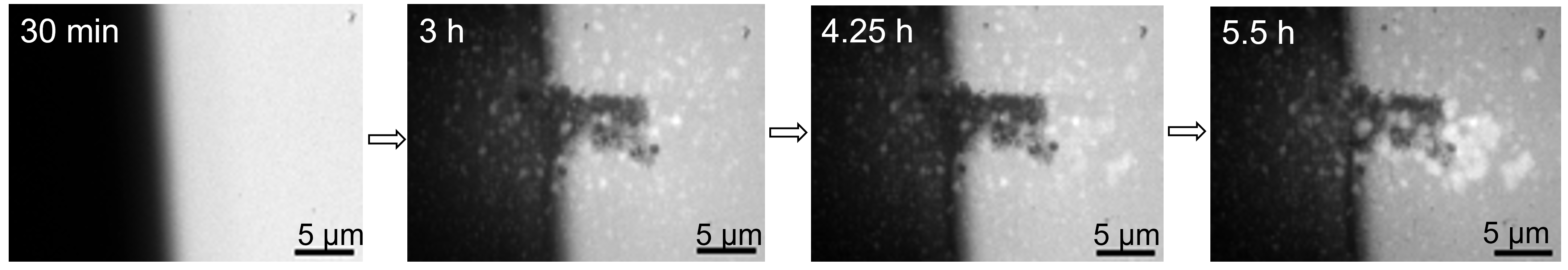
In situ cells are available for gas environments, supporting a temperature ramp of up to 1100 ℃, and for liquid environments, with temperatures of up to 80 ℃. With a resolution of 50 nm on the beamline, and sub-nanometre resolution on the TEM, we can thus perform an in situ multi-length scale analysis. Results from this setup so far include an in situ study of a catalytic system under redox reactions [1] and an investigation of iron corrosion in water at elevated temperatures (Figure 1).
[1] Parker et al., (2022) J Synchrotron Radiation, 29(2)
Figure 1: XRF images, Fe-Ka signal, of iron metal film exposed to brine solution, imaged on the I14 beamline using the in situ liquid setup. XANES measurements are performed in between the XRF measurements, in the area that is outlined in the 3h panel by a rectangle of white dashed lines, thus this area has been exposed to a higher X-ray dose. This demonstrates that the corrosion reaction is accelerated by the effects of the X-rays.
- References
Parker et al., (2022) J Synchrotron Radiation, 29(2)