Multi-technique, in-situ analysis workflow for battery materials in the scanning electron microscope
- Abstract number
- 171
- Presentation Form
- Poster & Flash Talk
- DOI
- 10.22443/rms.mmc2023.171
- Corresponding Email
- [email protected]
- Session
- Correlative and Multimodal X-ray Microscopy
- Authors
- Alexandra Stavropoulou (1), Dr Lucia Spasevski (1), Dr Joshua Lea (1), Kim Larsen (1), Dr Ute Schmidt (2), Dr Matthew Hiscock (1), Dr Dan Haspel (1)
- Affiliations
-
1. Oxford Instruments Nanoanalysis
2. WITec
- Keywords
batteries, anodes, cathodes, separators, battery materials, particle analysis
- Abstract text
The pressure for the development of green energy technologies is higher than ever. Batteries are required to store more energy and perform reliably for longer and under a range of conditions - often extreme heat or cold weather - while delivering power for demanding applications. Currently, the most prevalent battery technology is that of Li-ion batteries due to the outstanding energy and power density it offers, especially for demanding applications such as electromobility. Battery materials analysis is not only essential for understanding materials behaviour, failure mechanisms, and performance but also for designing better materials for future batteries. Here, we focus on in-situ scanning electron microscope (SEM)-based analytical techniques for the characterisation of battery materials.
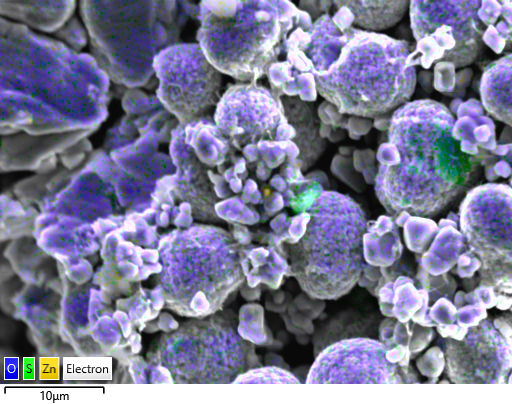
Quality assurance/quality control (QA/QC) of cathode precursor powders helps in cutting costs and saving expensive high-purity material resources by detecting contamination in powders (Figure 1) in a timely manner. The benefits that come with early contamination detection range from short/mid-term to long-term: 1) improvement of standard operating procedures by identifying the contamination source and 2) focusing on the battery as a finished product, reduction of the malfunction probability or premature failure owing to the presence of contamination particles which can pierce through the different components of a battery and, consequently, trigger short circuiting. QA/QC particle analysis is reliable and fast as it can be done with SEM-energy dispersive spectroscopy (EDS). Moreover, it is quite common in manufacturing for cathode precursor powder batch testing. Similarly, contamination can be found in anode materials.
Figure 1 ZnS contamination (in green) in NCM811 cathode precursor particles.
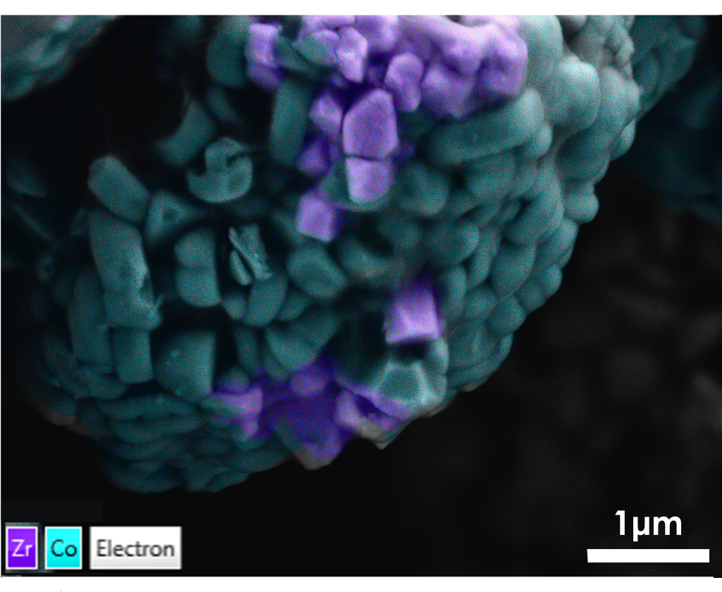
Apart from QA/QC, formulation testing is also key in battery materials analysis. One way of improving battery materials performance can be the addition of dopant elements. Dopants modify the material’s properties aiming to achieve resistance to degradation (cracking) and suppression of dendrite formation caused by cycling. High resolution SEM-EDS is a straightforward way to detect the dopant distribution on the particles surface (Figure 2). The next step of this workflow is qualitative mapping with wavelength dispersive spectroscopy (WDS). SEM-WDS can be complementarily used to quantify the dopant concentration, providing thus additional reassurance in checking the presence of the dopant element(s) even at very low concentration.
Figure 2 ZrO dopants (in purple) on NCM811 particle surface, mapped with windowless EDS detector.
Precursor cathode powders as well as cathodes can be characterised on the basis of their structural properties (grain size, texture). The electron image on its own does not provide enough information on the particle internal structure, especially because the cathode particles are made of elements with close atomic number (Z). As a result, their grey levels are also close and difficult to discern. However, this problem can be overcome by performing electron backscatter diffraction analysis (EBSD). SEM-EBSD band contrast maps clearly show the individual grains and their internal structure. EBSD also provides textural information (grain boundaries, size, shape, orientation) which can be used for failure analysis (cracking development and propagation) as well as material behaviour with cycling.
The incorporation of the RISE (Raman-SEM) system delivers a complimentary approach to battery analysis by offering a fully confocal optical stage within the SEM chamber. This provides additional molecular information and is easily integrated into the SEM analysis workflow. Such analysis is particularly beneficial when assessing the graphitic and amorphous phases of anode materials as changes after cycling can be distinguished. Furthermore, Raman mapping can identify the polymer separator materials meaning that structural changes due to charging and cycling can also be differentiated and closely monitored.
The aforementioned techniques (including Raman spectroscopy for SEM) come with the advantage that they can be performed in situ, in the same SEM chamber. Thus, data is collected under the same conditions in a single experiment. Apart from the workflow efficiency achieved, this is also of great importance for beam and/or air sensitive samples, the chemistry and structure of which can be easily altered (and the subsequent analysis of which can lead to errors). Additionally, data collected with such a multitude of techniques can be correlated and materials properties can be understood in greater depth.
- References