Correlative synchrotron X-ray techniques to study dental enamel remineralisation using self-assembling peptides
- Abstract number
- 100
- Presentation Form
- Poster & Flash Talk
- DOI
- 10.22443/rms.mmc2023.100
- Corresponding Email
- [email protected]
- Session
- Correlative and Multimodal X-ray Microscopy
- Authors
- Miss Reham Gonnah (2, 1), Dr Julia Parker (1), Professor Maisoon Al-Jawad (2), Dr Robert Davies (2)
- Affiliations
-
1. Diamond Light Source
2. University of Leeds
- Keywords
Correlative X-ray techniques
X-ray diffraction
X-ray tomography
Materials
Biological tissues
Dental Enamel
- Abstract text
This study focuses on utilising correlative X-ray techniques on the DIAD beamline at Diamond Light Source (DLS) to better understand the role of a soft material (self-assembling peptide (SAP) P11-4) in remineralising dental enamel. Combining X-ray tomography with X-ray diffraction mapping allowed visualisation and quantification of how the SAP affects mineral density as well as the texture and lattice parameters of reformed crystallites.
Dental caries is the most common problem presented in the clinic. Recently, the desire to shift from invasive restorative approaches to minimally invasive ones to treat the condition has increased. A soft material, namely SAP P11-4, has been studied as a potential treatment for early caries. It has been proposed to self-assemble in one dimension to form a network of fibres, which act as a scaffold for de novo hydroxyapatite nucleation. Nonetheless, detailed investigations of its mechanism of action/how it promotes enamel remineralisation are yet to be carried out in order to potentially optimise it for the treatment of early caries. Our study focuses on utilizing X-ray diffraction mapping and X-ray tomography simultaneously on the DIAD beamline at DLS to study the role of SAP P11-4 in remineralising dental enamel. A static experiment has been carried out on the beamline where the two techniques were used to scan the previously prepared enamel samples. The effects of SAP P11-4 on enamel remineralisation have been investigated by analysing the changes in texture and orientation of the hydroxyapatite crystallites, lattice parameters, and the mineral density with treatment.
Human teeth with sound enamel were sectioned to produce samples that were 2mm wide and 1mm thick. A 1mm x 1mm window on the enamel sample was created using acid-resistant nail varnish. Control samples were left untreated, while the rest of the samples were placed in demineralisation solution for 96 hours to create artificial lesions. Samples were remineralised for 120 hours with and without treatment with SAP P11-4. X-ray tomography and X-ray diffraction mapping were carried out on the DIAD beamline at DLS, and the mineral density values were extracted from the reconstructed image stacks after calibration using scanned phantoms. The azimuthal spread of the (0 0 2) hydroxyapatite diffraction peak (FWHM) was used to assess the amount of texture in the enamel, while Rietveld Refinement of the integrated diffraction patterns was carried out to extract the unit cell lattice parameters across the mapped regions.
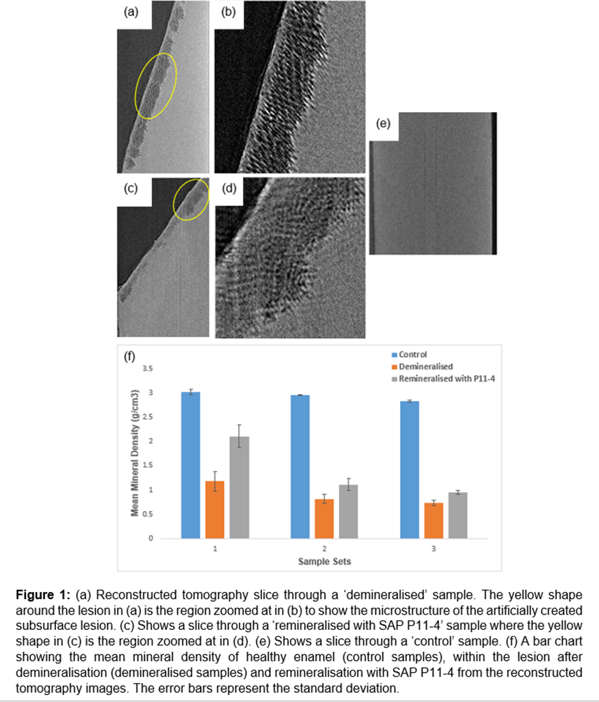
The X-ray imaging data show that the mean mineral density within the lesion decreases with demineralisation as expected, and increases in the samples treated with SAP P11-4 as shown in Figure 1f. Additionally, treatment with P11-4 is shown to change the microstructure of the enamel as shown in Figure 1 (b, d).
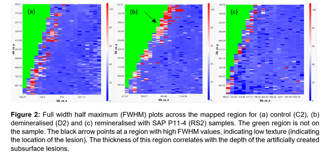
As shown in Figure 2b, the demineralised sample displayed a higher FWHM, for the (0 0 2) azimuthal peak, on the surface where the lesion was observed which indicates lower texture. This is expected as with demineralisation, the crystallites’ arrangement is disrupted, resulting in the disordering of the enamel crystallites. With P11-4 treatment, the FWHM is shown to increase on the surface, indicating higher texture (better ordering of the crystallites) as shown in Figure 2c. This indicates that the P11-4 could restore the alignment of the reformed crystallites through guided remineralisation rather than random deposition of calcium phosphate. The hydroxyapatite a and c lattice parameters were also found to decrease in regions that had been remineralised after treatment with P11-4, again suggesting that the SAP is influencing the remineralisation process.
The combination of multiple X-ray techniques can be used to better study the effect of soft materials on the microstructure and nanostructure of mineralised tissues such as dental enamel and bone. Using correlative techniques not only allows us to investigate the effects of soft materials on multiple length scales to build a full picture, but it also allows us to quantify different parameters such as mineral density, orientation and alignment of reformed crystallites, as well as the effect on the lattice parameters.
- References