Multimodal imaging of placenta histopathological specimens
- Abstract number
- 148
- Presentation Form
- Poster & Flash Talk
- DOI
- 10.22443/rms.mmc2023.148
- Corresponding Email
- [email protected]
- Session
- Multimodal Microscopy
- Authors
- Ms Georgia Mappa (4), Dr Nicolas Orsi (3), Dr Tiehan Shen (5), Dr Michele Cummings (3), Ms Harriet Pyrah (2), Ms Clare Freer (3), Mr Richard Oliver (1), Dr Pika Miklavc (5), Dr Huda Alzahrani (5)
- Affiliations
-
1. University of Leeds
2. University of Leeds/St James Teaching Hospital
3. University of Leeds/St James's Teaching Hospital
4. University of Leeds/St. James's Teaching Hospital
5. University of Salford
- Keywords
placenta histopathology, polarimetric microscopy, amyloids, multimodal microscopy
- Abstract text
Getting a better understanding of the placental origins of pregnancy complications is a first step towards developing novel prophylactic therapeutic interventions in obstetrics (e.g. reorchestrating pregnancy zone protein activity in the clearance of misfolded proteins) and improving both maternal and neonatal outcomes. The current study aims to validate the use of quantitative polarimetric microscopy in a histopathological context to gain an understanding of the histoarchitectural localisation of misfolded amyloid-β (Aβ) deposits in placentas from both normal and pre-eclamptic pregnancies, and to quantify the polarimetric characteristics of Aβ deposits. In turn, these will be related to placental integrity, function and disease presentation (including onset, severity and association with intra-uterine growth restriction).
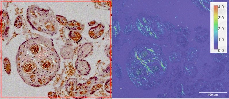
Recently, we have demonstrated the operation of a prototype photoelastic modulator-based polarimetric microscope developed in-house. In the case of liquid crystal droplets [1], different structures and the handedness of the internal molecular arrangements were readily observable. In the present work, we report a preliminary polarimetric imaging study of full thickness placental sections. Representative results are illustrated in Figure 1. The left image was obtained using a Congo red stained pre-eclamptic specimen and cross-polariser microscopy, where putative amyloid deposits showed a hint of ‘apple green’ birefringence, a standard clinical histopathological approach in the identification of suspected Aβ deposits. The same area was imaged using our polarimetric methodology, where the colour coding is in birefringent retardation in the unit of nm (right image). The regions of interest were clearly visible, and the birefringence strength was quantifiable by polarimetric imaging. The nature of these deposits is currently being confirmed by orthogonal means using immunohistochemistry and immunofluorescent imaging.
While this polarimetric methodology was readily applicable to histology specimens, it is also inherently integrable with other modern light microscopy imaging techniques. In this regard, we plan to explore bimodal fluorescence-based imaging in the same prototype microscope.
Figure 1: A sample image of a placenta stained with Congo red with cross-polariser microscopy in which putative amyloid deposits show a hint of ‘apple green’ birefringence (left). The same area imaged using our polarimetric methodology (right). The colour coding is in birefringent retardation in nm.
- References
[1] Gou, J., Shen, T.H., Bao, P. et al. A stokes polarimetric light microscopy view of liquid crystal droplets. Sci Rep 11, 16329 (2021). https://doi.org/10.1038/s41598-021-95674-4