Crystalline orientation mapping at 1kV using the eCHORD approach for increased spatial resolution.
- Abstract number
- 108
- Presentation Form
- Poster
- DOI
- 10.22443/rms.mmc2023.108
- Corresponding Email
- [email protected]
- Session
- Poster Session One
- Authors
- Dr. Clément Lafond (1), M. Thierry Douillard (3), Dr. Hassan Saad (2), Dr. Sylvain Deville (4), Pr. Sylvain Meille (3), Pr. Philippe Steyer (3), Dr. Sophie Cazottes (3), Dr. Gabriel L'Hôte (3), Dr. Cyril Langlois (3)
- Affiliations
-
1. CEA
2. CNRS-Saint-Gobain CREE,
3. INSA Lyon
4. Université Claude Bernard Lyon 1
- Keywords
Advances in SEM, Crystaline Orientation Mpping, Low Dose/Low Voltage Imaging and analysis, Electron Channeling Contrast
- Abstract text
Mapping of crystalline orientations obtained by scanning electron microscopy (SEM) using the Electron Back Scattered Diffraction (EBSD) technique is crucial to obtain local information on the microstructure of polycrystalline materials (grain size, texture, special grain boundaries, phase discrimination...). However, the spatial resolution is limited by the interaction volume of the electrons under the sample surface and the particular geometry of EBSD experiments (70° sample tilt). Because it is hardly possible to obtain routinely usable information in EBSD with an electron beam of energy lower than 5keV, some submicron features of the microstructure may not appear in the EBSD maps.
However, electron channeling contrast is still present at much lower voltages [1]. Actually, this contrast is the signal used by CHORD, an innovative approach to orientation mapping based on the variation of the backscattered electron signal during sample rotation [2]. To explore the possibility of obtaining orientation maps at very low voltages and thus with potentially increased spatial resolution, CHORD experiments using a primary beam acceleration of 1kV were performed on alumina samples with a bio-inspired lamellar structure mimicking the nacre of shellfish.
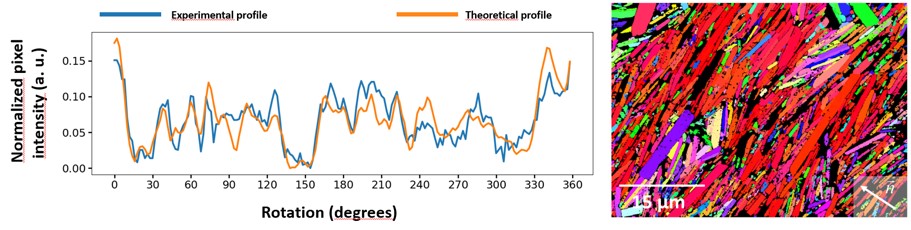
A series of 180 images (1024x768) with a frame time of 10s per image was acquired, with an acceleration voltage of 1kV, a tilt of 15°, and a 2° step of sample rotation between each image. For each pixel in the area of interest, a profile of backscattered intensity as a function of rotation is extracted, which is compared to a database of theoretical profiles to find the crystal orientation. It appears that the correspondence between an experimental profile and its theoretical counterpart is good enough to ensure a robust indexing, even at such a low acceleration voltage, as shown in Figure 1a. The full orientation map was then calculated (Fig. 1b), showing the same local texture information as the EBSD on the same sample.Figure 1 : on the left, correspondance example between CHORD intensity profiles, theoretical (orange) and experimental (bleu) ; on the right, associated orientation map.
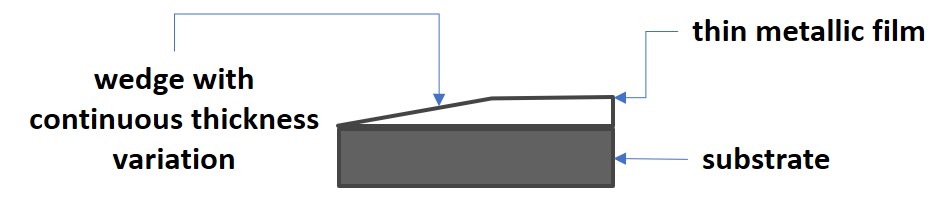
The gain in spatial resolution when working at such low accelerating voltage was then studied experimentally. CHORD and EBSD Experiments were carried out on metallic thin films on which a low-angle wedge has been milled from the surface of the film to the monocrystalline substrate using a Focused Ion Beam, providing a surface with controlled thickness variations (see Fig. 2).Figure 2 : sample design to obtain a controlled thickness variation surface: a wedge milled in a thin film deposited on a monocrystalline substrate using a focused ion beam.
For EBSD, the interaction depth under the surface was evaluated for different accelerating voltages by monitoring the substrate contribution in Kikuchi patterns for different film thicknesses along the wedge. For CHORD, orientation maps for differents acccelerating voltages have been acquired on the wedge surface. The fit quality between experimental intensity profiles and their theoretical counterpart is used as a marker to evaluate the interaction depth for the channeling contrast.
This preliminary result paves the way to the study of samples sometimes difficult to characterize by conventional EBSD due to the submicron characteristics of the microstructure (orientation gradients, fine precipitation...).
[1] Mikmeková, Š., Yamada, K., Noro, H. TRIP steel microstructure visualized by slow and very slow electrons. Microscopy, 62, (2013) 589–596.
[2] Lafond C., Douillard T., Cazottes S., Steyer P. and Langlois C. Electron CHanneling ORientation Determination (eCHORD): An original approach to crystalline orientation mapping. Ultramicroscopy, 186, (2018) 146-149.- References
[1] Mikmeková, Š., Yamada, K., Noro, H. TRIP steel microstructure visualized by slow and very slow electrons. Microscopy, 62, (2013) 589–596.
[2] Lafond C., Douillard T., Cazottes S., Steyer P. and Langlois C. Electron CHanneling ORientation Determination (eCHORD): An original approach to crystalline orientation mapping. Ultramicroscopy, 186, (2018) 146-149.