X-ray Ptychography Imaging of Human Chromosomes After Low-dose Irradiation
- Abstract number
- 251
- Presentation Form
- Poster Flash Talk + Poster
- DOI
- 10.22443/rms.mmc2021.251
- Corresponding Email
- [email protected]
- Session
- Poster Session 2
- Authors
- Ms Archana Bhartiya (3, 5, 4), Dr Darren Batey (2), Dr silvia cipiccia (2), Dr Xiaowen Shi (1), Dr Christoph Rau (2), Prof Stanley Botchway (4), Dr Mohammed Yusuf (3, 4, 6), Prof Ian Robinson (3, 4, 7)
- Affiliations
-
1. Department of Physics,New Mexico State University
2. Diamond Light Source
3. London Centre for Nanotechnology,University College London
4. Research Complex at Harwell
5. Department of Chemistry, University College London
6. Centre for Regenerative Medicine and Stem Cell Research,Aga Khan University
7. Condensed Matter Physics and Materials Science Division, Brookhaven National Lab
- Keywords
X-ray microscopy, Karyotype, Irradiation, Chromosome structure, Mass determination
- Abstract text
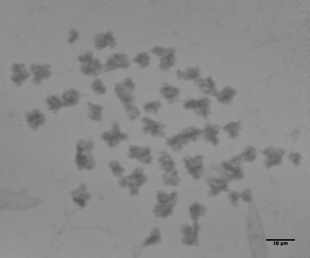
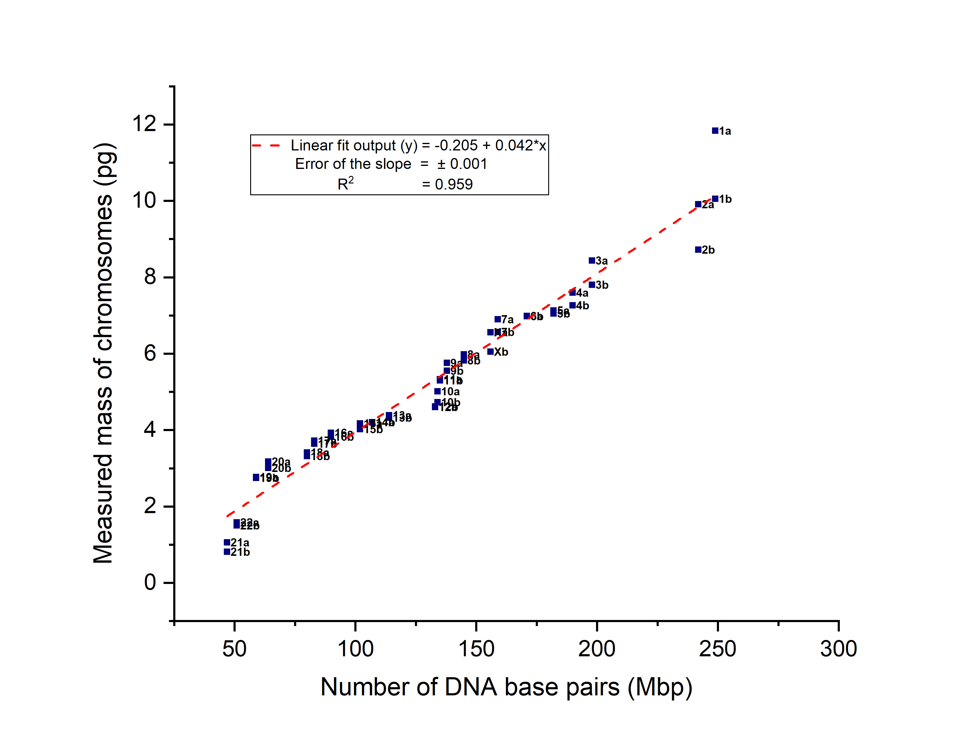
Studies into investigating the structure and composition of human chromosomes in the field of cytogenetics have spanned over several decades. In this work, we take advantage of the coherent X-rays available at the synchrotron facility at Diamond Light Source, Didcot, UK to extract individual masses (including DNA content and associated chromosomal proteins) of all 46 chromosomes. The experiments were performed, using the phase-sensitive X-ray ptychography method at the I-13 beamline. We have produced ‘X-ray karyotypes’ of unstained chromosome spreads (Fig.1) to determine the gain or loss of genetic material upon low-level X-ray irradiation doses due to radiation damage [1]. Two observations were made: first, the mass of each chromosome is 6-times higher than its theoretical DNA content. First, this suggests the presence of possible unknown nucleoproteins contributing to the excess mass. Secondly, the mass of the chromosomes is seen to increase upon low-level irradiation, suggesting the onset of the cell cycle repair mechanism. We show that the X-ray ptychography technique is useful for determining the total genome mass of species at the different stages of the cell cycle without invasive staining as well as the effects of ionizing radiation on the genome.
a) b)
Figure 1: Ptychographic reconstructed unstained chromosome spread obtained from non-irradiated T cells, a) phase-retrieval image, FoV: 32 µm × 32 µm, scale bar = 10 µm, b) X-ray karyotype of the same spread, with a best-linear-fit.
[1] A. Bhartiya, D. Batey, S. Cipiccia, X. Shi, C. Rau, S.W. Botchway, M. Yusuf, and I.K. Robinson. “X-ray Ptychography Imaging of Human Chromosomes after Low-dose Irradiation”. Chromosome Research, (2021).
DOI : 10.1007/s10577-021-09660-7.
- References
A. Bhartiya, D. Batey, S. Cipiccia, X. Shi, C. Rau, S.W. Botchway, M. Yusuf, and I.K. Robinson. “X-ray Ptychography Imaging of Human Chromosomes after Low-dose Irradiation”. Chromosome Research, (2021).
DOI : 10.1007/s10577-021-09660-7.