Using Multimodal X-Ray Microscopy for Structural and Chemical Nanoscale Imaging
- Abstract number
- 369
- Corresponding Email
- [email protected]
- Session
- Stream 3: X-ray Microscopy: A Powerful Tool to Aid the Understanding of Structures in the Life and Physical Sciences
- Authors
- Dr Julia Parker (1)
- Affiliations
-
1. Diamond Light Source
- Keywords
X-Ray Microscopy, Nanoprobe, Biomineral
- Abstract text
X-Ray Microscopy is a powerful tool for measuring local variations in composition, structure and morphology with application across a wide range of fields from geological, archaeological and life sciences to materials science. Recent advances in instrumentation, focussing optics and new imaging methodologies, coupled with a scientific demand for higher spatial resolution, has led to the construction of a number of hard X-ray nanoprobes at synchrotrons worldwide providing a broad range of techniques and beam-sizes.
The Hard X-ray Nanoprobe, Beamline I14, at Diamond Light Source provides spatial mapping of elemental composition by X-ray fluorescence (XRF), speciation by X-ray absorption near edge spectroscopy (XANES spectro-microscopy), structural phase by nano-X-ray diffraction (nano-XRD) and phase or electron density through differential phase contrast (DPC) and ptychography, with a particular emphasis on spectro-microscopy. These applications, and the benefits of combining multiple techniques, will be highlighted using examples such as the mapping of metallic anti-cancer drugs within cells, and the structure and composition of battery particles.
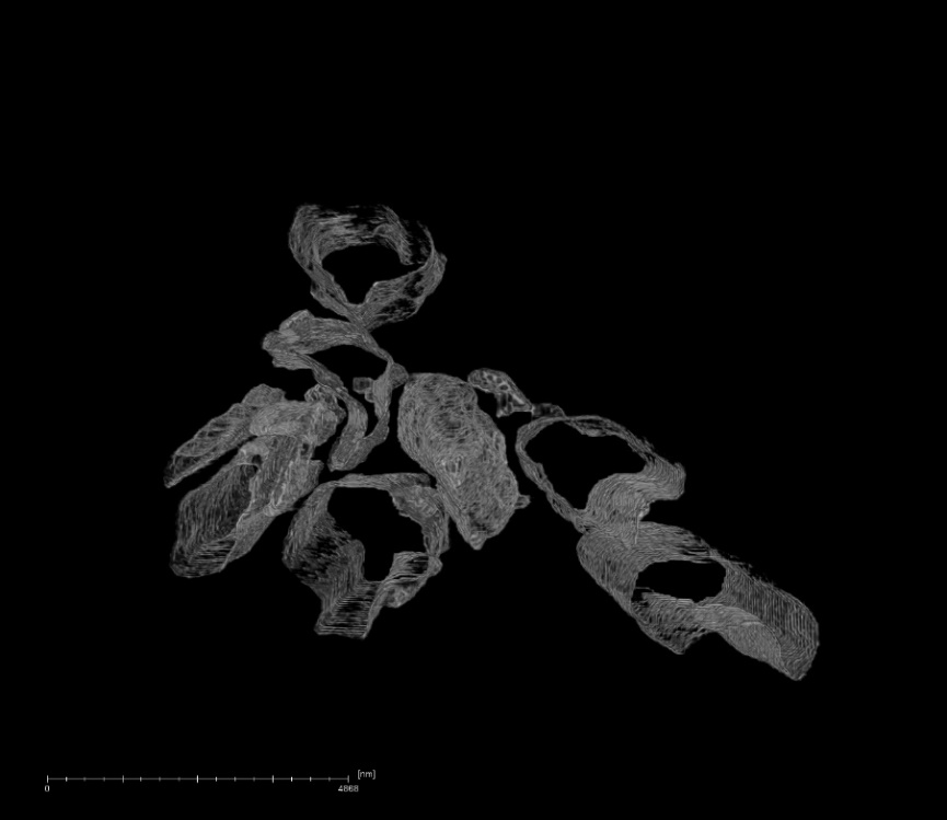
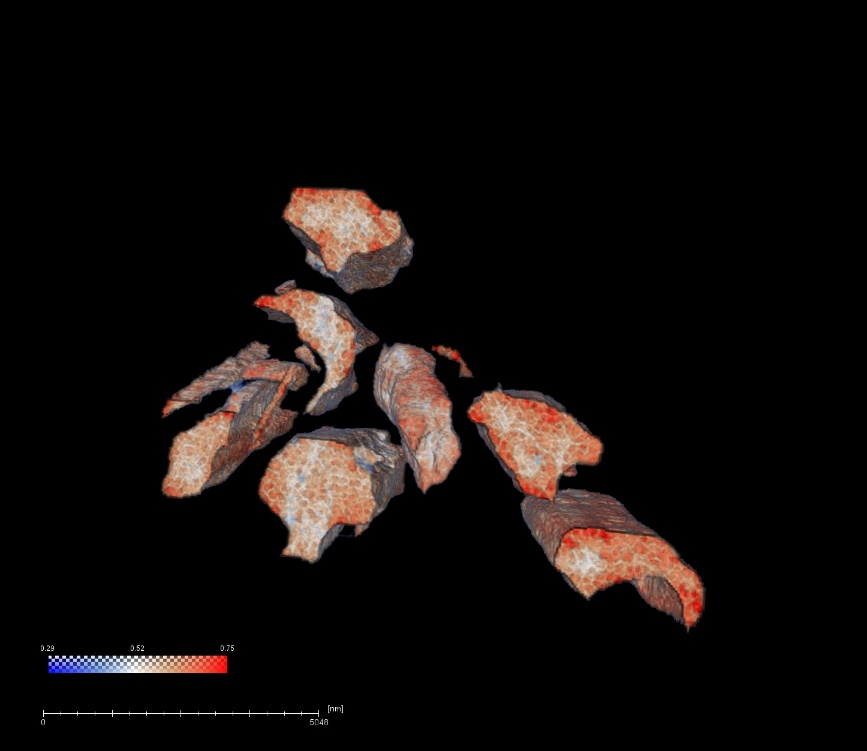
These advanced nano-imaging techniques also provide new opportunities to investigate the complex hierarchical structure of biominerals. Much research effort is being directed to discovering how mineralising organisms provide spatial, chemical, structural, textural and morphological control of inorganic mineral components, often creating unusual non-equilibrium crystal morphologies. Details of the underlying nanostructure of biominerals such as tooth, bone and shell, are essential in explaining their mechanical properties and offer clues to their formation mechanism. The potential is illustrated with, a ptychographic study of the calcite prisms from the shell of the mussel Mytilus Edulis (Figure 1.), underlining the role of the organic matrix in mineralisation, and a study of the mineralisation of collagen using multiple X-ray microscopy techniques, as a complement to electron microscopy, to gain insights to the mineralisation process across multiple lengthscales.
Figure 1. Ptychography reconstruction of M. Edulis calcite prisms showing the organic rich outermost 100nm (L) and denser mineral core (R)