The impact of strain on Z-contrast imaging of iron oxide nanoparticles
- Abstract number
- 226
- Presentation Form
- Submitted Talk
- DOI
- 10.22443/rms.mmc2021.226
- Corresponding Email
- [email protected]
- Session
- Stream 3: 3D+ Image Analysis
- Authors
- Mr Shuayl Alotaibi (2), Mr Toby Bird (2), Dr. Sitki Aktas (1), Dr. Leonardo Lari (3), Dr. Demie Kepaptsoglou (4, 2), Dr. Andrew Pratt (2), Prof. Roland Kröger (2)
- Affiliations
-
1. Department of Mechanical Engineering, Giresun University
2. Department of Physics, University of York
3. JEOL York Nanocenter
4. SuperSTEM
- Keywords
Z-contrast imaging
strain
Iron oxide nanoparticle
3D image
- Abstract text
It is well-established that mechanical, chemical and electronic properties of magnetic nanoparticles (MNPs), in particular metal and metal oxide NPs, are strongly affected by lattice strain1,2,3,4. Also, strain modifies the electronic surface structure of NPs, affecting the catalytic performance. Of particular interest are core/shell NPs with two different components constituting core and shell. Such shell can be the oxide forming on the surface of a metallic NP during exposure to air. This oxidation process is influenced by the strain induced by the oxide having a different lattice structure compared to the core1. Therefore, it is of great importance to understand strain in reactive NPs on the atomic level. A quantification of strain helps to understand the oxidation mechanism in metal oxide NPs and reveal whether lattice or the grain boundary diffusion dominate5. Our focus lies on the study of cubic iron-based magnetic nanoparticles with high-resolution aberration-corrected Transmission Electron Microscopy (TEM) using Z-contrast imaging in scanning TEM (STEM). This imaging method provides detailed information on atomic column positions and allows for a direct interpretation of image contrast in terms of variations in iron atom densities in the beam direction. In a previous publication, it was shown that the oxide shell reveals a large variation of strain associated with the oxide/oxide boundaries and hence we are interested in obtaining a better understanding of the impact of strain in oxides on the contrast variations in high annular angular dark field (HAADF) images. Previous studies show that strain due to point or extended defects has a significant impact on the contrast in Z-contrast images6,7 and our aim is to systematically study the correlation between volume strain and image intensity.
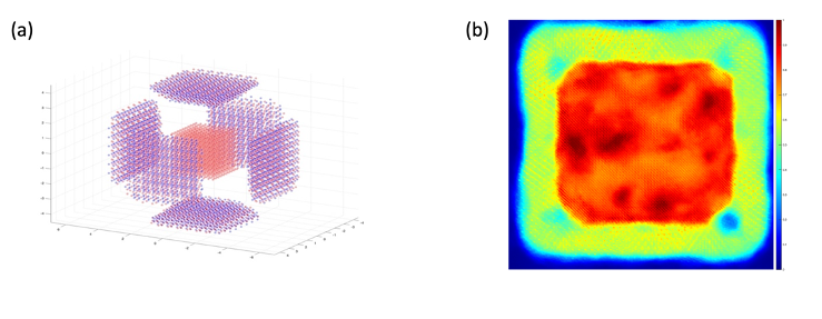
Figure 1a shows an idealized presentation of the principle components of a cubic oxidizing nanoparticle consisting of an iron core and six truncated pyramids which grow in size and shape upon progression of oxidation with Fig. 1b showing an experimental Cs corrected STEM image of an oxidised iron NP (color-coded).
Figure 1: (a) Idealised representation of the components of an oxidized cubic iron/iron oxide NP. Red: iron atoms, blue: oxide atoms. (b) False colored HAADF image of a core/shell Fe/Fe oxide nanoparticle.
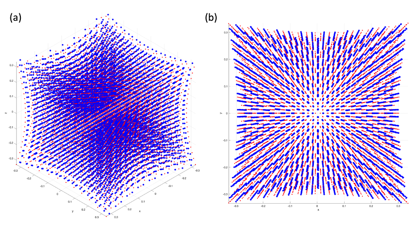
Figure 2: Gaussian strain field used in the simulations with a mean sigma value of 1.965 oriented along (a) [111] and (b) [001]. Red: iron, blue: oxide.
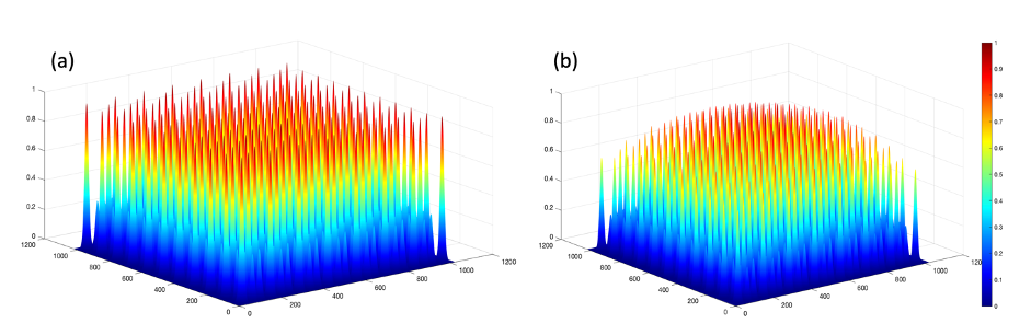
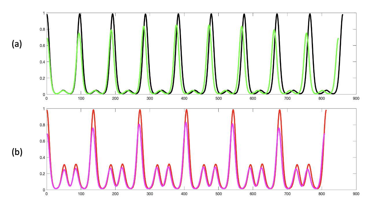
Although this does not represent a realistic scenario for strain distribution in oxidized iron nanoparticles, it allows for a systematic study of the atomic level response of the image intensity to strain. For image simulations we used the multislice imaging algorithm provided by the JEMS software8. A 3D gaussian strain profile with a maximum of 10% at the periphery of the NP was used in this model (Fig. 3). Line profiles taken across specific column positions (Fig. 4) were extracted and compared in Fig. 5. High intensity peaks correspond to Fe2+ columns and low intensity peaks to Fe3+ columns.
Figure 3: Intensities of Fe columns in the multislice simulated image of a magnetite NP in [011] zone axis orientation in the (a) absence and (b) presence of a gaussian strain profile with a maximum of 10% at the periphery.
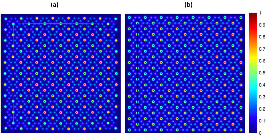
Figure 4: Multislice image simulation of a 6 nm magnetite cube in [011] zone axis orientation. (a) a strained and (b) unstrained magnetite nanoparticle. The simulation parameters: acceleration voltage = 200 kV, Cc = 1.2 mm, Cs = 0.01 mm, C5 = 10 mm, HAADF image, TDS = 0
Overall, the simulations reveal a twofold impact of strain: an expected shift of column positions with a column displacement and a spatially dependent reduction of column intensity. This intensity modulation is due to the displacement of individual atoms perpendicular to the beam direction with an effective reduction of scattering centre density in beam direction.
Figure 5: Line profiles taken along the column positions indicated in Fig. 4 for (a) an unstrained and (b) a strained magnetite NP. Green and black curves correspond to vertical and pink and red to horizontal profiles as indicated in Fig. 4. High intensity maxima correspond to Fe2+ columns whereas low intensity maxima correspond to Fe3+ columns.
Our studies show that small variations of strain have a significant impact on the intensity distribution in STEM images which need to be taken into account in the interpretation of high-resolution Z-contrast images in nanoparticles subjected to strain. This is particularly true for complex nanoparticles such as oxidized iron nanocubes, where strain is present at the interface between the iron core and the oxide shell as well as within the oxide shell and at the oxide grain boundaries leading to atomic displacements in beam direction and perpendicular to it.
- References
1. Gamler, J. T. L. et al. Effect of lattice mismatch and shell thickness on strain in core@shell nanocrystals. Nanoscale Adv. 2, 1105–1114 (2020).
2. Sneed, B. T., Young, A. P. & Tsung, C.-K. Building up strain in colloidal metal nanoparticle catalysts. Nanoscale 7, 12248–12265 (2015).
3. Chen, J. S., Liu, Y., Zhai, Y. & Fan, T. X. A new method to reliably determine elastic strain of various crystal structures from atomic-resolution images. Sci. Rep. 9, 16399 (2019).
4. Mavrikakis, M., Hammer, B. & Nørskov, J. K. Effect of Strain on the Reactivity of Metal Surfaces. Phys. Rev. Lett. 81, 2819–2822 (1998).
5. Pratt, A. et al. Enhanced oxidation of nanoparticles through strain-mediated ionic transport. Nat. Mater. 13, 26–30 (2014).
6. D.D. Perovic, C.J. Roussow, and A. Howie, Imaging elastic strains in high-angle annular dark field scanning transmission electron microscopy, Ultramicroscopy 52, 353 (1993).
7. P.J. Phillips, M. DeGraef, L. Kovarik, A. Agrawal, W. Windl, and M.J. Mills, Atomic-resolution defect contrast in low angle annular dark-field STEM, Ultramicroscopy 116, 47 (2012).
8. Stadelmann, P. JEMS - EMS java version. https://www.jems-swiss.ch/Home/jems/jemsv4_9731u2021.htm (2021).