Secondary Electron Hyper Spectral surface imaging for beam sensitive biomaterial characterisation
- Abstract number
- 270
- Presentation Form
- Submitted Talk
- DOI
- 10.22443/rms.mmc2021.270
- Corresponding Email
- [email protected]
- Session
- Stream 1: EMAG - Spectroscopy & Advanced SEM
- Authors
- Dr Nicholas Farr (2), Dr Antje Quade (1), Dr Jan Schäfer (1), Dr Cornelia Rodenburg (2)
- Affiliations
-
1. Leibniz-Institut für Plasmaforschung und Technologie e.V. (INP)
2. University of Sheffield
- Keywords
- argon plasma treatment, polymer characterization, polymeric bioma-terials, secondary electron emission, secondary electron hyperspectralgimaging
- Abstract text
Summary:
Is there electron microscopy spectroscopy method that can reveal local functional group variations at the nano- and micron scale on the surface of biomaterials? Such a technique would be transformative for biomaterial characterisation if it can be carried out with any sample preparation (e.g, thinning, coating etc). One of the most straightforward and widely used electron microscopes is the Scanning Electron Microscope (SEM) in the Secondary Electron (SE) imaging mode. Here we demonstrate SE Hyperspectral Imaging (SEHI) can be used to map functional groups involved in polymer cross-linking.
Introduction:
Biomaterials mechanical properties and surface functional groups are defined by their underlying chemical and structural relationships whose local variations determine cell growth propensity. For this reason, novel surface chemical spectroscopy and imaging methods on nano- and micro-scale levels are needed. In particular, the functional groups expressed and local variations in molecular order are major contributors to a biomaterials mechanical properties and consequently the biocompatibility of the material. Established averaging methods for the estimation of molecular order and chemical functional groups do not provide information of their spatial distributions across the biomaterials surface. Here we show that SEHI can provide the necessary spectral information and that can also be exploited for SE imaging. To show this we used SEHI to analyse poly(glycerol sebacate) methacrylate (PGS-M) surfaces resulting from the application of the industry standard autoclave sterilisation, to that of surfaces resulting from the use of low-pressure Argon glow discharge in order to explore Argon plasma as a model treatment on route to achieving a potential biomaterial sterilisation method.
Methods/Materials:
The PGS-M materials were either enclosed in a gas semi-permeable bag which was exposed to low-pressure argon (AR) glow discharge or placed into an Autoclave. Observation of the surface morphology of the resulting PGS-M surfaces was performed using a Scanning Electron Microscope (FEI Nova Nano 450 SEM). To avoid surface charging and consequent damage to the sample, a low accelerating voltage (1 KV) with typical vacuum pressure of 10−5 mbar at a working distance of 4 mm was applied. The FEI Nova Nano 450 SEM is provided with a through lens detector which includes a voltage controlled deflector electrode. The deflector electrode channels the signal into the SE detector. The deflector electrode is set to a predetermined number of deflector voltages and an image is generated for each deflector voltage. Spectra and hyperspectral images are acquired through post-processing of such image series [1].
Results and Discussion:
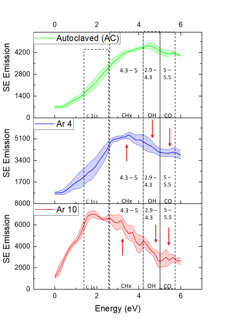
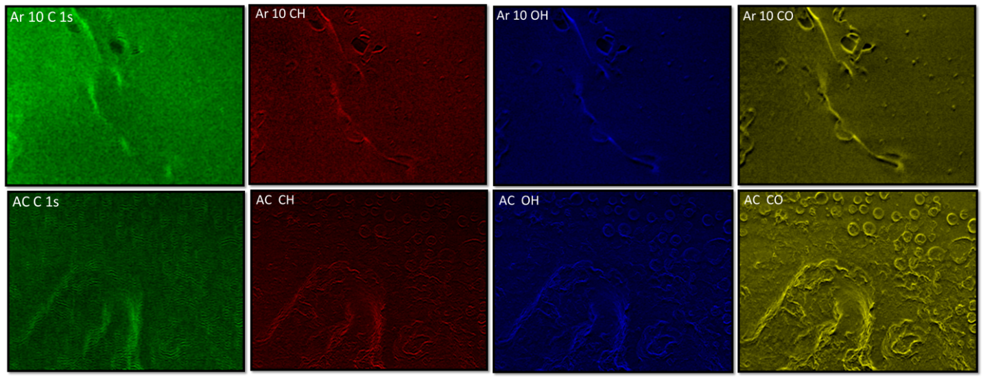
X-ray photoelectron spectroscopy (XPS) a well-established surface analysis tool was used on reference materials to identify energy ranges in the SE spectra associated with specific functional groups [2]. Figure 1 shows the SE spectra and resulting SEHI images of AC PGS-M, 10-minute Argon plasma treated PGS-M (Ar 10) and 4-minute Argon plasma treated PGS-M (Ar 4). Changes within the SE spectra for peak emissions associated with (CHx, OH and CO) chemical bonding were present within the different test samples. An example of SEHI ability to characterise chemical functional groups is given in the SE spectra for Argon plasma treated and AC samples observed emission within the region of 5-5.5 eV, related to C=O bonding. The spectra show that post Argon plasma treatment the emissions in the 5 – 5.5 eV range are greatly diminished. Argon plasma treatment is known to cleave away C-O-C bonds attached to the methacrylate within PGS-M. By cleaving away this bond, removal of methacrylate decreases the amount of C=O bonds present within the polymer. It is worth highlighting that the decrease in C=O bonding is most visible in the Ar 10 rather than Ar 4 samples which signifies that the cleavage of methacrylate units is time and region dependent. Such SE emission differences can then be mapped and it is evident when you compare Ar 10. CO SEHI chemical maps, with that of AC samples a decrease in CO emission is visible. SEHI findings were then corroborated with that of. SEHI therefore offers an efficient way to map functional groups with the required image resolution together with an ability to map at multi-length scales.
Conclusion:
We have demonstrated that using SEHI it is possible to map functional groups variations with ease in the scanning electron microscope. This work therefore proposes an innovative surface approach to providing researchers with effective tools to visualise and characterise novel biomaterials in the SEM.
Figure 1 -.A) Secondary electron spectra for AC, Ar 4 and Ar 10 treated PGS-M highlighting the regions identified as associated with functional group emissions. B) SEHI images generated from the component analysis of Ar 10 and AC. Mapping C 1s, CH, OH and CO bonding. Reproduced and rescaled from Farr N, Thanarak J, Schafer J, Quade A, Claeyssens F, Green N, et al. Understanding Surface Modifications Induced via Argon Plasma Treatment through Secondary Electron Hyperspectral Imaging. Adv Sci (Weinh). 2021;8(4):2003762 under the Creative Commons Attribution International License (CC BY) http://creativecommons.org/licenses/by/4.0/.
The authors thank EPSRC for funding under SEE MORE: Secondary Electron Emission‐Microscopy for Organics with Reliable Engineering Properties (EP/N008065/1), and studentship for N.F. (EP/R513313/1).
Keywords: argon plasma treatment, polymer characterization, polymeric bioma-terials, secondary electron emission, secondary electron hyperspectralimaging
argon plasma treatment, polymer characterization, polymeric bioma-terials, secondary electron emission, secondary electron hyperspectralimaging- References
[1] M. Dapor, R. C. Masters, I. Ross, D. G. Lidzey, A. Pearson, I. Abril, R. Garcia-Molina, J. Sharp, M. Unčovský, T. Vystavel, F. Mika, C. Rodenburg. Secondary electron spectra of semi-crystalline polymers–A novel polymer characterisation tool?. Journal of Electron Spectroscopy and Related Phenomena 222 (2018): 95-105.
[2] N. Farr, J. Thanarak, J. Schäfer, A Quade, F Claeyssens, N Green, C Rodenburg. Understanding Surface Modifications Induced via Argon Plasma Treatment through Secondary Electron Hyperspectral Imaging. Adv. Sci. 2021, 8, 2003762.