Polarization Mapping in Single Crystal Bulk Perovskite Oxides by Atomic Resolution STEM
- Abstract number
- 346
- Presentation Form
- Submitted Talk
- Corresponding Email
- [email protected]
- Session
- Stream 5: Late Breaking
- Authors
- Wanbing Ge (2), James Gott (2), Jonathan Peters (1), Marin Alexe (2), Ana Sanchez (2)
- Affiliations
-
1. Trinity College Dublin
2. University of Warwick
- Keywords
scanning transmission electron microscopy, perovskite, ferroelectric
- Abstract text
Ferroelectric materials are of great importance in the fields of high frequency electronics, non-volatile memories, microsystems and more1. Spontaneous polarizations inside ferroelectrics are divided into different domains and separated by domain walls. Measuring the displacement of cations and anions by means of electron microscopy is a key method to understand the origin of ferroelectricity and the behaviour of domains and domain walls at the atomic level2. While polarization mapping in ferroelectric perovskite oxides has been carried out concerning heavy A-site and B-site atoms, analysing the role of oxygen atoms is hindered as they are much lighter than their neighbours and are usually not distinguishable by conventional scanning transmission electron microscopy (STEM), although their importance in the formation of ferroelectricity has been well studied and modelled3,4.
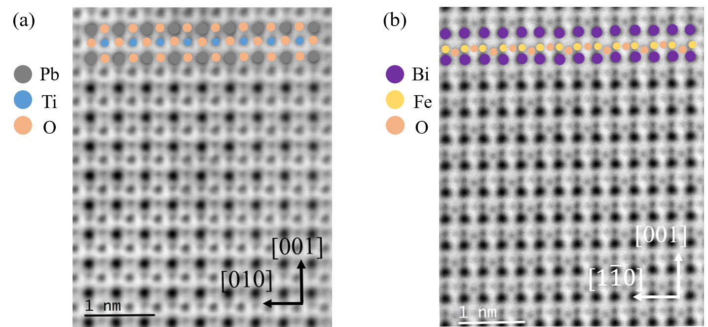
Here atomic resolution STEM imaging is used to determine the atomic position in ferroelectric perovskite PbTiO3 and BiFeO3 single crystals. Annular dark field (ADF) imaging is used to determine positions of heavy atoms like Pb or Bi, while annular bright field (ABF) imaging (Figure 1) provides position information of O atoms5. Specimen with different orientations have been prepared to give a thorough picture of the positions and displacement in real space. In PbTiO3, polarization is observed to point mainly up/down along the c axis and the oxygen atoms parallel/perpendicular to this direction have slightly different shift magnitudes. While for BiFeO3 the case is more complicated as the polarization is along the body diagonal of the rhombohedral unit cell (i.e. <111> direction), so we could only see the in-plane component of the polarization if the orientation of specimen is not perpendicular to it. The atomic shift of each atom can be visualized and the contribution to the total spontaneous polarization can be calculated. Domain patterns from the same crystal are also mapped by piezoelectric force microscopy (PFM) for a good comparison with STEM.
Figure 1. Atomic resolution ABF image of (a) PbTiO3 and (b) BiFeO3
- References
(1) Setter, N.; Damjanovic, D.; Eng, L.; Fox, G.; Gevorgian, S.; Hong, S.; Kingon, A.; Kohlstedt, H.; Park, N. Y.; Stephenson, G. B.; Stolitchnov, I.; Taganstev, A. K.; Taylor, D. V.; Yamada, T.; Streiffer, S. Ferroelectric Thin Films: Review of Materials, Properties, and Applications. J. Appl. Phys. 2006, 100, 051606. https://doi.org/10.1063/1.2336999.
(2) Jia, C.-L.; Nagarajan, V.; He, J.-Q.; Houben, L.; Zhao, T.; Ramesh, R.; Urban, K.; Waser, R. Unit-Cell Scale Mapping of Ferroelectricity and Tetragonality in Epitaxial Ultrathin Ferroelectric Films. Nat. Mater. 2007, 6 (1), 64–69. https://doi.org/10.1038/nmat1808.
(3) Shirane, G.; Pepinsky, R.; Frazer, B. C. X-Ray and Neutron Diffraction Study of Ferroelectric PbTiO 2. Acta Crystallogr. 1956, 9 (2), 131–140. https://doi.org/10.1107/S0365110X56000309.
(4) Kubel, F.; Schmid, H. Structure of a Ferroelectric and Ferroelastic Monodomain Crystal of the Perovskite BiFeO 3. Acta Crystallogr. B 1990, 46 (6), 698–702. https://doi.org/10.1107/S0108768190006887.
(5) Peters, J. J. P.; Apachitei, G.; Beanland, R.; Alexe, M.; Sanchez, A. M. Polarization Curling and Flux Closures in Multiferroic Tunnel Junctions. Nat. Commun. 2016, 7 (1), 13484. https://doi.org/10.1038/ncomms13484.