Optimal experiment design for characterising structures containing multiple types of elements using 4D scanning transmission electron microscopy
- Abstract number
- 151
- Presentation Form
- Poster Flash Talk + Poster
- DOI
- 10.22443/rms.mmc2021.151
- Corresponding Email
- [email protected]
- Session
- Stream 1: EMAG - 4D-STEM
- Authors
- Duygu Gizem Sentürk (1, 2), Annick De Backer (1, 2), Sandra Van Aert (1, 2)
- Affiliations
-
1. EMAT, University of Antwerp
2. NANOlab Center of Excellence, University of Antwerp
- Abstract text
Aberration corrected scanning transmission electron microscopy (STEM) has become a powerful technique for materials characterisation of complex nanostructures. In combination with quantitative methods, structural information such as number of atoms can be retrieved from experimental images. To obtain reliable atom-counting, one can predict the optimal experimental settings. For this purpose, the probability of error (Pe) concept was introduced [1,2] using statistical detection theory that enabled us to find the optimal detector collection regions to count the number of atoms from monotype nanosystems. To extend the problem for the investigation of complex hetero nanostructures, the use of pixelated detectors offers important advantages since it allows us to create conventional STEM images with any number of virtual detectors without the need for pre-configured fixed detector angles [3] or ultimately to use the full 4D STEM dataset.
In the present work, we have extended the concept of the Pe to investigate the potential benefit of analysing multiple 2D STEM images when counting atoms of different chemical nature. In this methodology, the atom-counting problem is formulated as a statistical hypothesis test, where each hypothesis corresponds to a specific number of atoms in an atomic column. The Pe corresponds to the probability to choose the wrong hypothesis. To compute the Pe, realistic 4D STEM simulations were performed from which multiple 2D STEM images were generated with varying inner and outer detector angles. From these 2D images, so-called scattering cross-sections (SCS) can be computed corresponding to the total scattered intensity for each atomic column. This measure has been shown to perform as an optimal criterion for atom-counting [2]. Moreover, the presence of electron counting noise is taken into account when computing the Pe.
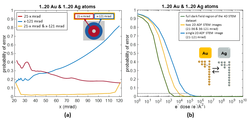
To illustrate the concept, the Pe has been calculated to distinguish between pure Ag and Au columns up to 20 atoms thick for SCSs for an incident electron dose of 104 e-/Å2. In Figure 1a, the red curve shows the evaluation of the Pe as a function of the outer angle of a single ADF detector with a fixed inner angle, while the blue curve shows the evaluation as a function of the inner angle with a fixed outer angle. From these results, it follows that the optimal Pe equals 16% when using a single ADF detector. The yellow curve represents the calculation of the Pe for the combination of scattering cross-sections extracted from two non-overlapping detectors. This clearly shows that the Pe can be reduced to only 2% when two sets of independent data are employed. In Figure 1b, the Pe is evaluated as a function of the electron dose under the optimal detector settings for a single ADF detector, for a combination of two optimal independent detectors, and for the full dark field region of the 4D STEM dataset. The dashed horizontal line shows that when aiming for a Pe as low as 2%, the electron dose can be further reduced to 103 e-/Å2 when exploring the 4D STEM dataset.
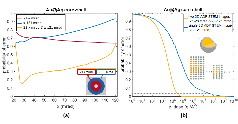
Subsequently, the research problem was extended to a Au@Ag core-shell nanoparticle to retrieve information about different types of elements in a binary mixed atomic column structure with minimum Pe. In this research problem, the total thickness of the Au-Ag mixed atomic columns varies from 1 up to 20 atoms and the core is assumed to be located in the centre. The Pe has been calculated to distinguish both the total number of atoms in the atomic column and the number of Au atoms in the core. In Figure 2a, the Pe is evaluated as a function of the detector angles for an incident electron dose of 104 e-/Å2 resulting in a higher Pe as compared to the previous study for pure columns up to 20 atoms thick. However, in this case, the Pe can be reduced from 65% to 15% when a combination of two ADF detectors is employed rather than a single optimal ADF detector. In Figure 2b, the evaluation of the Pe as a function of the incident electron dose is shown. The dashed horizontal line shows that to reach a Pe of 2%, the electron dose can be reduced by a factor of 103 when using two optimal non-overlapping detectors. Consequently, the required electron dose to count the number of atoms for different types of elements in a binary mixed atomic structure can significantly be lowered with the use of two independent 2D STEM images reconstructed from a 4D STEM dataset.
In summary, we have shown that the concept of the Pe can be used to find optimal strategies to count atoms from 4D STEM datasets. In particular, the method is generalised to create a set of multiple 2D STEM images providing independent information concerning thickness, composition, and ordering of the atoms along the viewing direction [4].
Figure 1: Pe to distinguish between pure Ag and Au columns (a) as a function of the outer angle of a single ADF detector with fixed inner angle (red), the inner angle of a single ADF detector with fixed outer angle (blue), and the common angle x for two independent detectors (yellow) for an incident electron dose of 104 e-/Å2 (b) Pe as a function of the incident electron dose calculated for a single ADF detector (blue), two independent ADF detectors (yellow), and for the full dark field region of the 4D STEM dataset (green).
Figure 2: Pe for Au@Ag core-shell nanoparticle to distinguish between the total number of atoms and type of atoms in a binary mixed atomic column varying from 1 up to 20 atoms (a) as a function of the outer angle of a single ADF detector with fixed inner angle (red), the inner angle of a single ADF detector with fixed outer angle (blue), and the common angle x for two independent detectors (yellow) for an incident electron dose of 104 e-/Å2 (b) Pe as a function of the incident electron dose calculated for a single ADF detector (blue) and two independent ADF detectors (yellow).
- References
[1] J. Gonnissen et al., Appl. Phys. Lett. 105 (2014) 063116
[2] A. De Backer et al., Ultramicroscopy 151 (2015) 46
[3] N. Shibata et al., J. Electron Microsc. 59 (2010) 473
[4] This work was supported by the European Research Council (Grant 770887 PICOMETRICS to SVA and Grant 823717 ESTEEM3). The authors acknowledge financial support from the Research Foundation Flanders (FWO Belgium) through project fundings and a postdoctoral grant to ADB.