Matching algorithms for elemental quantification and few tilt tomography in 2D materials
- Abstract number
- 289
- Presentation Form
- Submitted Talk
- Corresponding Email
- [email protected]
- Session
- Stream 1: EMAG - 2D Materials
- Authors
- Dr. Christoph Hofer (2), Dr. Viera Skákalová (3), Jonas Haas (1, 4), Prof. Jannik Meyer (1, 4), Prof. Timothy Pennycook (2)
- Affiliations
-
1. Eberhard Karl University of Tübingen
2. University of Antwerp
3. University of Vienna
4. Natural and Medical Sciences Institute at the University of Tübingen
- Keywords
2D materials, Quantitative STEM, 3D imaging
- Abstract text
Introduction
Identifying the position and chemical identity of each atom in a specimen is the ultimate goal of structural characterization. With the rise of aberration correctors in scanning transmission electron microscopy (STEM) the Z-contrast based annular dark-field (ADF) imaging technique even allows to distinguish light elements in single layer materials [1]. However, residual aberrations are difficult to manually detect and correct under the low doses needed for beam sensitive materials hampering the analysis of ADF images. Moreover, a conventional tomographic approach for three-dimensional imaging is difficult due to the requirement of a large number of projections [2]. I will introduce the approach of using matching algorithms to overcome these difficulties.
To perform quantification of images that is robust to the presence of both residual aberrations and noise, algorithmic matching of simulated and experimental data can be used [3]. A simulation is performed from an initial guess. Successive simulations are then iteratively optimized to match the experimental data as well as possible by adjusting the aberrations and atomic positions used in the simulation. Once convergence is achieved, the atomic scattering factors are finally included in the optimization.
Matching algorithms also enable tomography with a much reduced number or tilt angles. I will present results illustrating 3D structure determination of 2D materials such as graphene [4] directly from just two tilt angles. This is indeed sufficient as long as each atom can be identified individually in each projection and the connectivity matrix can be obtained showing which atom is which in the comparison of each view. Under these circumstances, a very similar optimization process is realized where the model is matched so that the simulated projections in each tilt angle are matched to the corresponding experimental data set. The reconstruction is demonstrated using the ADF signal, but is also expected to work with any other signal providing atomic-resolution.
Materials & methods
ADF STEM measurements were conducted using a Nion UltraSTEM100 operated at 60~kV and a JEOL JEM-ARM200F operated at 80 kV at a convergence angle of ~30 mrad, and tilts separated by ~20 deg. Ptychography data will be collected using a microsecond dwell time capable custom camera at EMAT.
Results and Discussion
Analysis reveals that our novel intensity method achieves more reliable results compared to other quantification methods in the presence of small non-round aberrations. The method even allows us to extract quantitative atomic intensities if the aberrations are strong. As a specific example, this approach allows to quantify different light-elements in graphene in the presence of a slight three-fold astigmatism which wouldn’t have been possible via other established approaches.
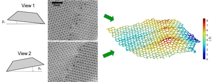
The three-dimensional analysis reveals significant deformation of defect sites in 2D materials. Specifically for graphene grain boundaries, a correlation between the deformation amplitude and the misorientation angle is demonstrated. Figure 1 shows one example of a reconstructed grain boundary using two atomically resolved STEM images captured at tilt angles separated by 20 degrees. The analysis also enables one to study single-atomic out-of-plane dynamics, which we have demonstrated for impurities in graphene.
We will discuss the extension of the few tilt tomography method to thicker 2D materials such as transmission metal dichalcogenides, and the addition of simultaneous ptychographic data to the ADF signal to facilitate the simultaneous location of both heavy and light atoms.
The dose-efficiency of ptychography should also allow a lower dose to be used for such tomography. Alternatively, the dose efficiency can be used to image at more tilts for a given dose budget. With more tilts it becomes easier to handle materials that are thicker than a single atomic layer. In order to further improve the reliability of the reconstruction, a hybrid approach is introduced, where the energy of the model is included into the optimization.
Conclusions
Our new methods provide reliable intensities from atomically resolved STEM images. They allow one to decouple intensity variations arising from aberrations, beam tails and varying bond distances from those arising from sample variations. For light-element samples, they allow sharp intensity distributions around single elements to be obtained. They also allow the three-dimensional structure of graphene to be obtained showing that atomically thin materials are far from being flat, especially at defect sites. An extension of the algorithm to a wide range of 2D materials is further discussed.
Figure 1: Two-tilt reconstructed graphene grain boundary showing a significant out-of-plane deformation induced by the line defect
- References
[1] Krivanek, O. et al., Nature 464, 571–574 (2010).
[2] A. C. Kak et al., IEEE Engineering in Medicine and Biology Society, New York: IEEE Press (1988).
[3] Hofer C. et al., submitted
[4] Hofer C. et al., 2D materials 5, 045029 (2018)
[5] Research funded by the Ministry of Science, Research and Art Baden-Württemberg and by the European Research Council (ERC) under the European Union’s Horizon 2020 research and innovation programme via Grant agreement No. 802123-HDEM.