Investigation of life science samples using an annular silicon drift detector at low beam currents
- Abstract number
- 201
- Presentation Form
- Poster Flash Talk + Poster
- DOI
- 10.22443/rms.mmc2021.201
- Corresponding Email
- [email protected]
- Session
- Poster Session 2
- Authors
- Max Patzschke (1), Dr Nils Schlüter (2), Dr Andrew Menzies (1)
- Affiliations
-
1. Bruker Nano
2. Museum für Naturkunde
- Keywords
Sea urchin, low kV, analysis, diatome, microbial mat, FlatQUAD, life science, annular silicon drift detector, shadow effects, Bruker, SDD, XFlash® FlatQUAD, ultra low beam current, SEM, EDS,
- Abstract text
Silicon drift detectors (SDD) have become state of the art technology in the field of EDS microanalysis. The accuracy and precision which SDDs offer is comparable or better than WDS (Ritchie et al., 2012; Cubukcu et al., 2008). Special detector designs and concepts have immensely improved the performance of SDDs and their applications to complex samples.
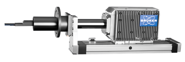
The XFlash® FlatQUAD (Fig. 1) has been developed for applications that are limited by normal EDS, for example, shadow effects and low count rates when applying low beam currents. The XFlash® FlatQUAD is inserted between the pole piece and the sample, and ideally suited for the analysis of topographically complex samples. Shadowing effects are minimized by collecting X-rays using four separate detector segments. This detector covers a very large solid angle (1.1 sr) and allows sufficient data collection at low beam currents on beam sensitive samples. Even at beam currents in the pA range, a sample can be investigated without carbon coating under high vacuum. Compared to low vacuum analysis, this results in higher spatial resolution of the SEM images and X-ray element mappings.
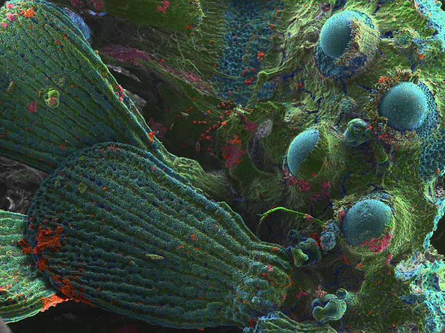
In this study a sea urchin (Fig. 2) and a microbial mat of diatoms were investigated without the requirement of any sample preparation.
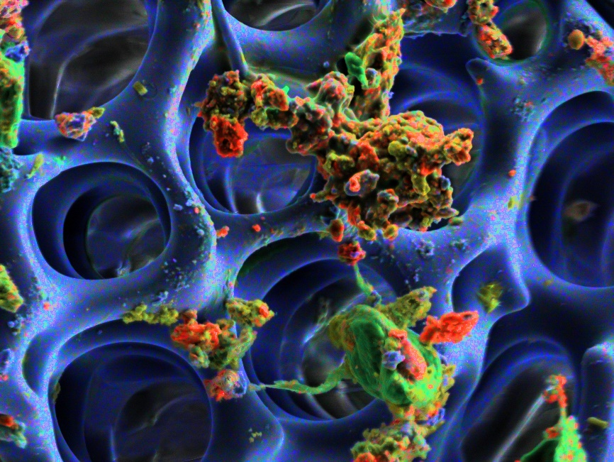
The EDS map shows a sea urchin (Paracentrotus lividus) collected in the Aegean sea near Athens, Greece. The bumps on the right are the sockets where the urchins long spines are attached. They are being held in place by muscles and collagen so that they can be moved if the urchin wants to hide in a crevasse and they can be held rigid in defense mode, e.g. when we step on it. In the center of the micrograph individual sand grains are discernable in red. The skeleton (Figure 3) of the sea urchin is produced by biomineralization, consisting of high-magnesium calcite.
The architecture of the skeleton reveals a sponge-like lightweight structure (Figure 3), which can bare extremely high weights. Such information is only possible with the annular design, avoiding shadowing and collecting highest count rates at low beam currents.
For element distribution mappings of complex structures like these and other beam-sensitive life science samples, the XFlash® FlatQUAD, is unmatched.
Figure 1: The XFlash® FlatQUAD
Figure 2: Sea urchin
Figure 3: Skeleton of the sea urchin
- References
Ritchie et al., 2012;
Cubukcu et al., 2008