Elucidation of short range order, defects and beam sensitivity in Ca2MnO4 Ruddlesden-Popper oxides through Transmission electron microscopy
- Abstract number
- 81
- Presentation Form
- Poster
- DOI
- 10.22443/rms.mmc2021.81
- Corresponding Email
- [email protected]
- Session
- Poster Session 4
- Authors
- Mr. Satyam Choudhury (1), Professor Rajiv Mandal (1), Dr. Joysurya Basu (1)
- Affiliations
-
1. Department of Metallurgical Engineering, Indian Institute of Technology (BHU)
- Keywords
Ruddlesden-Popper, Electron diffraction, Modulation, Defects
- Abstract text
Two dimensional Ruddlesden-Popper (RP) layered perovskite oxides are well known for their functional application as functional materials and in the field of alternative energy. Can+1MnnO3n+1 based RP compounds have layered arrangement of n octahedral blocks intercalated between rock salt layers. These multi-layered arrangement of conducting octahedra with insulating rock salt type layers within different family members (n = 1, 2, 3) leads to complex electro-magnetic transport behaviours. Therefore, this class of RP phases exhibit giant colossal magneto resistance behaviour and thermoelectric properties [1-2]. The motivation for this work is to illustrate short range ordering within nano regime, underlying defects, and stability of the phase in question under electron beam over prolonged exposure, primarily through electron diffraction and diffraction contrast imaging (DCI).
CaCO3 and Mn2O3 were mixed thoroughly in 4:1 molar ratio by weight then the mixture was converted into pellets which are placed inside alumina crucible to carry out heat treatment through solid state route. Crucible containing pellet was placed at 850°C inside muffle furnace for 24 hours then it was annealed to room temperature. The pellets were grounded, then were pelletized again and were kept inside muffle furnace at 1100° C for 14 hours. Finally it was annealed to room temperature.
Bulk characterization of synthesised phases were carried out through X-ray diffraction (XRD). It is observed that the d-spacings and relative intensities of peaks match mostly with that of the compound Ca2MnO4 reported earlier as tetragonal phase with space group I41/acd and lattice parameter a = 5.183Å and c = 24.117Å by Takahasi et al. during 1993 [3]. The structure of Ca2MnO4 has been simulated through VESTA and well correlated with experimental XRD data. Statistical sampling of this phase had been carried out through transmission electron microscopy [4]. Selected area electron diffraction (SAED) pattern confirms the doubling of lattice parameter along c-axis due to counter-tilt of consecutive MnO6 octahedra. The structure of tetragonal phase is confirmed through systematic tilting electron diffraction experiments. Finer tilting (<0.05°) about particular zone axis reveals the change in distribution of satellite spots revealing complex short range ordering. Diffraction signature of commensurately modulated unit has been observed. Signature of forest dislocation where assembly of dislocation entangled each other was observed through DCI. Fault in stacking sequence was quantified by correlating SAED pattern and DCI. Investigating particle was exposed to electron beam for prolonged period of time. Crystalline to amorphous phase transition has been captured. Detailed structural analysis of the phase will be presented.
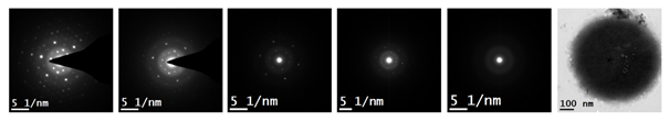
Figure 1 – Illustrate gradual drop of diffracted intensity upon continuous exposure to electron beam at 200 KV till amorphization.
- References
[1] Bendersky et al., (2003). Transmission electron microscopy study of Ruddlesden–Popper Can+ 1MnnO3n+ 1 n= 2 and 3 compounds. Journal of Solid State Chemistry, 174(2), 418-423.
[2] Azulay et al., (2020). Enhanced Charge Transport in Ca2MnO4-Layered Perovskites by Point Defect Engineering. ACS Applied Materials & Interfaces, 12(44), 49768-49776.
[3] J. Takahashi and N. Kamegashira, (1993). X-ray structural study of calcium manganese oxide by rietveld analysis at high temperatures [Ca2MnO4.00]. Materials research bulletin, 28(6), 565-573.
[4] D. B. Williams and C. B. Carter, (2009). Transmission Electron Microscopy, vol. 5, no. 721.
[5] The author would like to acknowledge the support from Department of Science & Technology inspired FIST programme.