Electron ptychographic imaging of polymer and macromolecular ordering
- Abstract number
- 112
- Presentation Form
- Poster
- DOI
- 10.22443/rms.mmc2021.112
- Corresponding Email
- [email protected]
- Session
- Poster Session 4
- Authors
- Mr. Botao Hao (1), Prof. Hazel Assender (1), Prof. Peter Nellist (1)
- Affiliations
-
1. University of Oxford
- Keywords
4D STEM, electron ptychography, polymer, macromolecule
- Abstract text
Polymers and macromolecules are widely used in the medical and pharmaceutical fields due to their outstanding biocompatibility and biodegradability, low density, and relatively low cost. However, the lack of microstructural study on these materials limits the ability to tailor them for broader and deeper applications.
As important methods for microstructural research, electron microscopic techniques have the potential to image polymers and macromolecules, but they are challenging for conventional electron microscopies because of their low crystallinity, poor contrast and severe beam sensitiveness [1]. Therefore, an advanced electron microscopic technique is required for the microstructural study. Electron ptychography makes use of 4D STEM data, and hence has lower sample damage and better phase-contrast compared to traditional techniques, which is helpful to overcome the existing challenges [2]. Here we apply electron ptychography to explore the fine structures of polyvinyl alcohol (PVA, a polymer) and amphiphilic peptides (macromolecules) respectively.
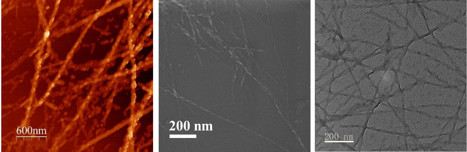
By CTEM, AFM and SEM, the morphology of peptides was firstly studied since previous research demonstrated the connection between the morphology and structure [3]. As shown in Figure 1, peptides exhibit a fibrous morphology, and the thickness of the fibres will change with the concentration of the peptide and the pH of solutions.
Figure 1. AFM (left), SEM (mid) and TEM (right) images of a peptide, FEFEPKFKF at pH 7 with 1 mg/mL in DI water
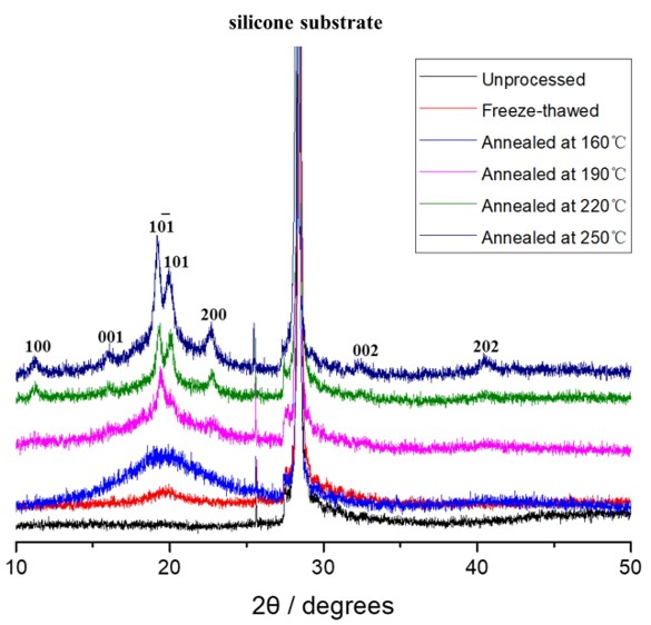
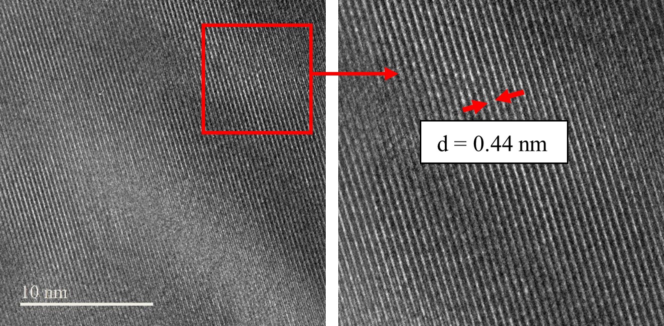
Then PVA and peptides were freeze-thawed and annealed for the higher crystallinity for electron ptychographic study. XRD, FTIR and HRTEM were subsequently applied to confirm the effects of freeze-thawing and annealing and get the basic crystal information. Figure 2 shows these methods do crystalise the PVA, and the increase of annealing temperature further facilitate the crystallinity of PVA. Furthermore, and a typical d-spacing of PVA (at its 101 plane) at 0.44 nm was acquired by HRTEM.
Figure 2. XRD scheme of PVA
Figure 3. HRTEM images of PVA
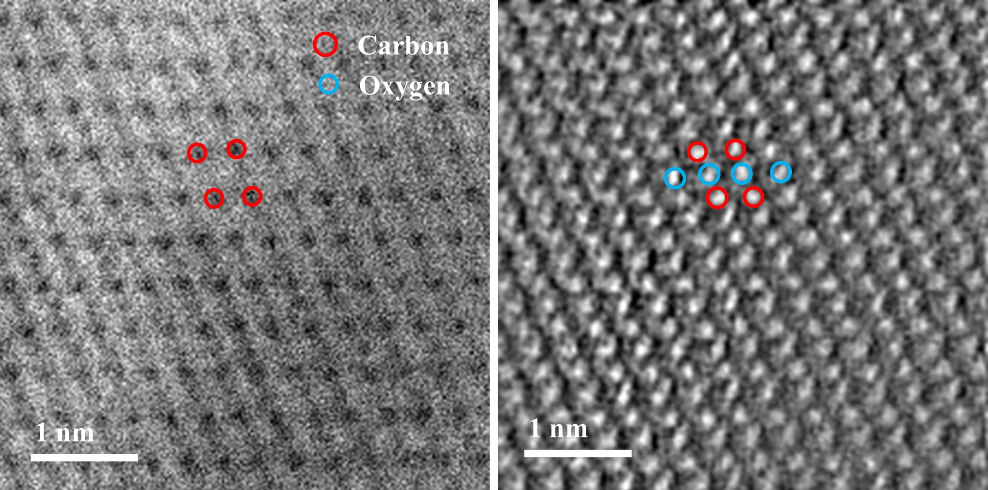
After these, electron ptychographic data were acquired with the aid of the aberration-corrected STEM and the pixelated 4D STEM detector. Electron ptychographic data were finally reconstructed by Single-side band (SSB) and Wigner-distribution deconvolution (WDD) methods. The reconstructed atomic-resolution images can show how the light atoms order in the materials. As shown in Figure 4, for the PVA sample, both carbon and oxygen atoms of the material can be clearly viewed after reconstruction, and in comparison, only carbons are visible in conventional STEM methods.
Figure 4. ABF STEM (left) and SSB reconstructed ptychographic (right) images of PVA
In summary, this project explores the application of electron ptychographic technique in polymers and macromolecules. To date, we have managed to study the morphology of peptides, increased the crystallinity of peptides and PVA, and got a clear view of PVA atomic structure via electron ptychography. In the future, the methodology can be expanded to other polymeric and macromolecular systems, and thus, better microstructural understandings could contribute to better medical and pharmaceutical applications of polymers and macromolecules.
- References
[1] Sawyer, L., et al. (2008). Polymer microscopy.
[2] Yang, H., et al. (2016). Nature Communications, 7, 12532.
[3] Roberts, D., et al. (2012). Langmuir, 28(46), 16196-16206.