Design of electron ptychography experiments through simulations
- Abstract number
- 338
- Presentation Form
- Poster Flash Talk + Poster
- DOI
- 10.22443/rms.mmc2021.338
- Corresponding Email
- [email protected]
- Session
- Stream 1: EMAG - 4D-STEM
- Authors
- Dr. Mohsen Danaie (2), Dr. Darren Batey (1), Dr. Thomas Slater (2), Dr. Christopher Allen (2, 3)
- Affiliations
-
1. Diamond Light Source Ltd.
2. Diamond Light Source Ltd., electron Physical Science Imaging Centre (ePSIC)
3. University of Oxford
- Keywords
ptychography, simulation
- Abstract text
With the wider accessibility of fast direct-electron detectors, collecting electron diffraction patterns in the scanning mode of the transmission electron microscope is becoming common place. These datasets – also known as 4D-STEM, with two dimensions on the scan array and two on the detector plane containing the diffraction corresponding to a given probe position – can be used to reconstruct both the complex object and the probe using ptychographic algorithms. Solving for the object via ptychography, in principle, can relax the requirements on the optical hardware to achieve atomic resolution imaging1 and has also been shown to be beneficial in reducing the total dose received by the sample2. Given the large number of parameters that the experimentalist operating a modern electron microscope can vary, carrying out an electron ptychography experiment, while ensuring that the collected data is conducive to a satisfactory reconstruction, can at times be a daunting task. Here we present a workflow to simulate a matrix of optical and sampling conditions to provide feedback on the optimal conditions to be used for the electron ptychography experiment. As there are various ptychographic reconstruction algorithms with different sampling criteria, our focus here is the extended ptychographical iterative engine (ePIE) implemented by one of the present authors in the ptyREX python library3.
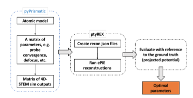
The workflow used in this study is presented in the schematic shown in Figure 1. The atomic model corresponding to the phase under investigation is imported to the Prismatic simulation package in python, and a multislice simulation is performed with the 4D-STEM output saved4. Python interface allows systematic variation in the input parameters of the optics such as probe convergence semi-angle, probe aberrations and also the probe step size in the simulated data. This data is then modified to incorporate noise and electron dose and fed into the ptyREX reconstruction algorithm. Following the ePIE reconstructions we then evaluate the outcomes based on a set of quantitative metrics. This allows us to identify the optimal experimental parameters for a given set of metrics used.
Figure 1. Workflow for evaluating a set of parameters in an electron ptychography experiment.
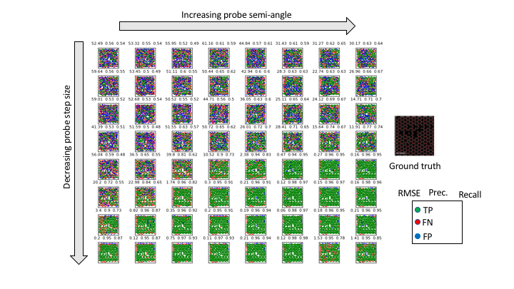
As a case study, we looked at a graphene atomic model with some defects included in the structure. With the probe defocus set to result in roughly the same probe diameter in each case, we varied the sampling in the real space and diffraction discs overlap in reciprocal space, by changing the probe step size and the probe convergence semi-angle, respectively. The reconstruction outcome was then evaluated with comparison to the projected potential of the initial model. True positive (TP) cases were atomic positions in the reconstruction matched with one in the model, false negative (FN) cases defined as missing atoms in the reconstruction and false positives (FP) as atoms in the reconstruction where no atom is present in the model. These cases are presented with different colours in Figure 2 matrix. We used three quantitative metrics: (1) root-mean-squared error of the distances between the identified versus the known atomic positions, (2) Precision: TP / (FP + TP), and (3) Recall: TP / (FN + TP). Using these, we identify the optimal experimental parameter in this case as 50 mrad probe convergence angle with 80% real space probe overlap. This is close to what we have found experimentally at 80 kV.
Figure 2. The outcome of ePIE reconstruction for the simulated matrix of ptychographic data. The ground truth used for evaluation along with a legend of the matrix is shown on the right-hand side.
We are now exploring the robustness of ePIE reconstructions to the potential experimental errors in various input parameters. This would help identify the key critical parameters that require closer scrutiny for successful experiment. Our aim is to have this workflow implemented such that it is user-friendly as much as possible to provide Users at ePSIC a working framework to better design their electron ptychography experiments.
References
1. J. Rodenburg and A. Maiden in P.W. Hawkes, J.C.H. Spence (Eds.), Springer Handbook of Microscopy, 2019.
2. J. Song, C. S. Allen, S. Gao, et al. Sci Rep 9, 3919 (2019).
3. D. J. Batey, Ptychographic Imaging of Mixed States, Thesis, University of Sheffield (2014).
4. C. Ophus, Advanced Structural and Chemical Imaging 3(1), 13 (2017).