Application of unsupervised machine learning methods to scanning electron nanobeam diffraction (SEND) data: case study for domain mapping in P2 and O3-type Sodium-Ion Battery cathode material
- Abstract number
- 311
- Presentation Form
- Submitted Talk
- Corresponding Email
- [email protected]
- Session
- Stream 4: Diamond Light Source Session 1
- Authors
- Mr Andy Bridger (3, 2, 1), Professor Bill David (2, 3), Dr Mohsen Danaie (1), Dr Keith Butler (2), Dr Thomas Wood (2)
- Affiliations
-
1. Diamond Light Source
2. STFC
3. University of Oxford
- Keywords
Unsupervised Machine Learning, Domain Mapping, Cluster Analysis, Structure Analysis, Structure Determination, Energy Materials, SEND Data Processing
- Abstract text
This work aims to develop a workflow that combines both informed and data-driven approaches to condensing the dimensionality of the electron diffraction data. Using these ‘latent space’ representations of the scanned region, we can apply clustering algorithms to identify regions of similar character. By combining the results of multiple autonomous approaches, we would be able to map domains within the crystal and associate a level of confidence to given classifications. Incorporating this as a pre-processing unsupervised workflow would drastically improve the ability to characterise the nano-scale structure of materials, both through the production of significantly signal-boosted diffraction data and reduction in the laborious manual investigation.
With the advent of fast direct-electron detectors, capturing large scan arrays of electron diffraction data is becoming more and more accessible. The scanning electron nanobeam diffraction technique lends itself well to microscope automation and can cover areas in the micrometres length scale and hence, while still capturing microstructural details not accessible via powder x-ray diffraction (PXRD), provides statistically significant insight into the crystallographic defects and domain morphology in the microstructure. Larger probe sizes and lower magnifications typically used in SEND also result in reduced electron dose on the sample, compared to atomic-resolution STEM, making this technique suitable for beam-sensitive phases. The quantity of SEND data collected during a day on the microscope can run well above 1TB. This is far more information than can be comprehensively processed by hand within a reasonable time frame. As a case study, we are implementing this approach to study the complex microstructure that drives leading Sodium-ion battery materials.
The sodium-ion batteries we investigated are among the best worldwide with performances that are comparable with Li-ion batteries using LiFePO4 as cathode material. Our cathode materials are based on the same layered metal oxide structures as the commercial lithium NMC and NCA cathodes but have a more complex structural polymorphism. These structures have been variously labelled P3, O2, OP4, Z, Z’, O3’, O3’’ and O3’’’, however, these labelled phases rarely exist in any extended form as the intercalation process is largely stochastic. Comprehensive knowledge of the different structures is essential for our understanding of these materials and, due to the potential complexity of phase disposition, these materials lend themselves to our unsupervised domain mapping approach.
We have investigated a number of state-of-the-art unsupervised learning approaches to reduce the dimensionality of our datasets and to represent them on a lower-dimension manifold. For the SEND data collection, we aligned a JEOL Grand-ARM microscope at 300 kV with the probe corrector optics turned off, allowing us to reduce the probe convergence semi-angle to around 1 mrad. We collected the diffraction data on a MerlinEM quad detector (Quantum Detectors) with each dataset reshaping to 256x256 in the probe scanning plane and 515x515 on the diffraction plane.
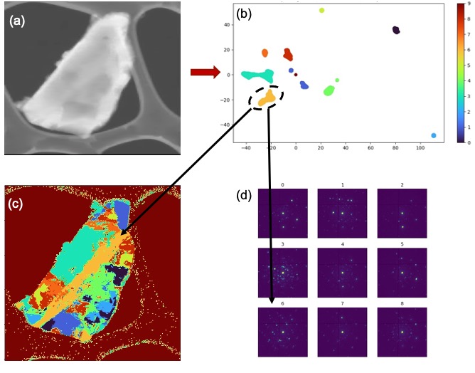
As an example, we present the results of this workflow, using a combination of Common Peak Selection (for initial dimensionality reduction with minimal information loss)– t-distributed Stochastic Neighbour Embedding (for further non-linear dimensionality reduction and cluster separation) and Gaussian Mixture Model (for clustering the data). This approach produced a domain map with plausible phase dispositions as seen in Figure 1.
Figure 1 – (a) the original unmapped scan region, (b) the two-dimensional representation of the key peak data reduced using a t-distributed Stochastic Neighbour Embedding (t-SNE) and clustered with a Gaussian Mixture Model (GMM) (note that the cluster at [0,0] corresponds to the uncomputed background regions was added afterwards for completeness), (c) a domain map produced from the clusters present in the manifold, (d) the composite diffraction patterns pertaining to the identified regions
The incoherence of some of the composite diffraction data along with the busier regions in the map, highlight the utility that will be provided by comparing multiple complementary domain maps to improve the confidence of the classification. A multi-prong approach is advantageous as any individual low dimensionality representation will result in a loss of information (the extent of this depends on the ‘inherent’ dimensionality of the data) and so using multiple representations allows us to combine different domain maps with different intrinsic information retained in order to segment the data in a way that still reflects all the information initially available.

