4D in-situ microtomography and image analysis of aerosol filtration
- Abstract number
- 210
- Presentation Form
- Poster Flash Talk + Poster
- Corresponding Email
- [email protected]
- Session
- Stream 4: Diamond Light Source Session 2
- Authors
- Mr Matthew Jones (4), Dr David Eastwood (4), Dr Andrew York (2), Dr Timothy Hyde (2), Dr Malte Storm (1), Prof Sarah Haigh (3)
- Affiliations
-
1. Diamond Light Source
2. Johnson Matthey
3. University of Manchester
4. University of Manchester at Harwell
- Keywords
X-ray Microtomography, Microscopy, In-situ, Aerosol, Filtration, 4D imaging
- Abstract text
Understanding the microscale processes underlying filtration is fundamental to optimising filter design and understanding aging. We have developed and integrated a novel in-situ flow rig for the 4D imaging of aerosol filtration in vehicle particulate filters at Diamond Light Source beamline I13-2. This combined imaging and aerosol flow system has revealed the local microstructure at positions where particulates are captured and agglomerate, allowing us to assess the factors affecting filter performance.
Gasoline particulate filters (GPFs) are used in current and next generation gasoline vehicle exhausts in order to meet environmental regulations that restrict the amount of toxic particulate matter (PM) emitted (Joshi & Johnson, 2018). Thus, these filters are a key technology in effectuating a cleaner transition to electric transport. GPFs are wall flow filters constructed of macro-porous cordierite extruded into long channels. In use the exhaust aerosol flows down these channels and through the porous walls of the cordierite, depositing PM on the wall surfaces and in the internal pore space of the filter. These deposits impede gas flow through the filter, increasing backpressure and reducing the fuel efficiency of the engine. Herein lies the key trade off in efficient GPF performance; low backpressures that improves fuel efficiency verses high filtration efficiency that prevents toxic substances entering the urban environment. Key to understanding this trade-off is observing the evolution of the pore network and the deposits within.
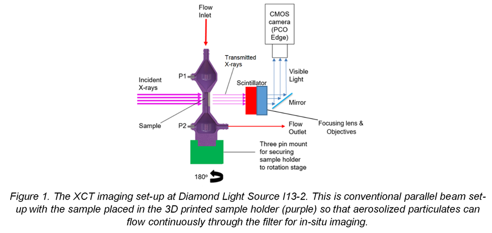
4D in-situ x-ray microtomography (XMT) allowed us to resolve the 3D microstructure of the filter, the deposited particulates, and importantly how the deposits evolve with time during a filtration process. We achieved this 4D study by creating a novel aerosol flow rig for in-situ parallel beam XMT. This consisted of an air compressor that provided head pressure to a dust generator which output a steady aerosol stream into the flow rig. We flowed TiO2 particles with nominal diameter 25nm, which displayed similar aerodynamic and clustering behaviours to real world engine exhaust. The aerosol then flowed through the customised 3D printed sample holder which maintained stability during tomographic rotation but was thin enough to minimize beam attenuation. Static pressure measurements were obtained before and after the filter position. The sample was placed at the thin central section of the holder, as shown below in Figure 1, such that the aerosol flowed through the centre of the filter.
Tomographic x-ray imaging used a ‘pink beam’ spectrum at beamline I13-2 (Pešić, 2013), combining multiple harmonic peaks from the undulator source. For fast imaging a projection was captured every 0.18o in a 180o fly scan with an exposure of 0.01s. The high flux from the synchrotron source gave sufficient time resolution to resolve the structure of the filter and deposit with only minimal change during each scan. We obtained 1 tomogram per minute, taking flat and dark field images at the beginning of each sequence of tomography scans, rather than after each scan, in order to minimize unnecessary movements. The imaging set-up is illustrated below in Figure 1.
Systematic analysis of the data used the following workflow: (1) A filtered back projection tomographic reconstruction. (2) Image pre-processing, including distortion correction, cropping, z-axis resampling, de-zinger, non-local means and Gaussian. (3) Binarization of the ‘zeroth’ volume (i.e. clean unloaded filter) into filter and void. (4) Registration of the subsequent deposit loaded volumes onto the zeroth volume. (5) Histogram rescaling across the entire 4D data set. (6) Masking using the ‘zeroth’ filter volume to mask-out the filter phase in the subsequent volumes, and (7) applying a global threshold to segment the deposit phase. Due to the large size of the processed data set (50 GB in 20 × 8 bit volumes of 1125 x 1125 x 2110 pixels) and the grey level overlapping of the phases of interest, this required a programmatic method using a range of python based software (Imageio, Scikit-image, Simple-ITK, OpenCV, and other packages within the Anaconda distribution).
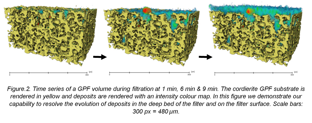
The resulting time resolved tomography is shown as volume renderings in Figure 2. We can observe the initial deposits in the internal deep bed pores of the filter growing between minutes 1 and 6. At 6 minutes we can see that the deposits in the internal wall have now ‘blocked’ the key flow paths through the filter and have become slightly denser as they have ‘filtered out’ aerosol flowing through the pore throat. Hence, after minute 6, deposits start forming primarily on the wall surface as shown in the minute 9 volume. This is the transition from deep bed to cake filtration as resolved through imaging, this coincided with the point at which we measured an increase in the backpressure from the manometer.
In conclusion 4D in-situ XMT allows us to observe the evolution of deposit position, extent and relative density with a time resolution of 1 tomogram per minute. We have achieved sufficient resolution to observe pore filling and pore blocking events and the transition from deep bed filtration to cake filtration. The data from this method has potential for model validation for computerised fluid dynamic based filtration models. This methodology highlights opportunities for studying the function of a range of operational particulate filters including machine intake filters, engine exhaust filters and respiratory filters.
- References
Joshi and Johnson (2018) - Joshi, A., Johnson, T.V. Gasoline Particulate Filters—a Review. Emiss. Control Sci. Technol. 4, 219–239 (2018). https://doi.org/10.1007/s40825-018-0101-y
Peši´c , (2018) - Peši´c, Z.D.; Fanis, A.D.; Wagner, U.; Rau, C. Experimental stations at I13 beamline at Diamond Light Source. J. Phys. Conf. Ser. 2013, 425